Aug 21 2010
For their look into the nanoworld, the Jülich researchers used a scanning tunneling microscope.
Its thin metal tip scans the specimen surface like the needle of a record player and registers the atomic irregularies and differences of approximately one nanometre (a billionth of a millimetre) with minuscule electric currents. However, even though the tip of the microscope only has the width of an atom, it has not been able so far to take a look inside molecules.
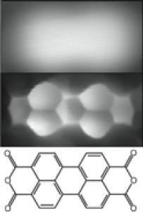
 The Juelich method makes it possible to resolve molecule structure where only a blurred cloud was visible before.
"In order to increase the sensitivity for organic molecules, we put a sensor and signal transducer on the tip," says Dr. Ruslan Temirov. Both functions are fulfilled by a small molecule made up of two deuterium atoms, also called heavy hydrogen. Since it hangs from the tip and can be moved, it follow the contours of the molecule and influences the current flowing from the tip of the microscope. One of the first molecules studied by Temirov and co-workers was the perylene tetracarboxylic dianhydride compound. It consists of 26 carbon atoms, eight hydrogen atoms and six oxygen atoms forming seven interconnected rings. Earlier images only showed a spot with a diameter of approximately one nanometre and without any contours. Much like an X-ray image, the Jülich scanning tunneling microscope shows the molecule's honeycombed inner structure, which is formed by the rings.
"It's the remarkable simplicity of the method that makes it so valuable for future research," says Prof. Stefan Tautz, Director at the Institute of Bio- and Nanosystems at Forschungszentrum Jülich. The Jülich method has been filed as a patent and can easily be used with commercial scanning tunnelling microscopes. "The spatial dimensions inside molecules can now be determined within a few minutes, and the preparation of the specimen is based predominantly on standard techniques," says Tautz. In the next step, the Jülich scientists are planning to calibrate the measured current intensity as well. If they are successful, the measured current intensities may allow the type of atoms to be directly determined.
After publishing initial images produced with the new method in 2008, the research group of Tautz and Temirov has now been able to explain the quantum mechanical principle of operation of the deuterium at the tip of the microscope. Their results were supported by computer-assisted calculations by the working group of Prof. Michael Rohlfing at the University of Osnabrück. The so-called short-range Pauli repulsion is a quantum-physical force between the deuterium and the molecule which modulates the conductivity and allows us to measure the fine structures very sensitively.
The Jülich method can be used to measure the structure and charge distribution of flat molecules which can be used as organic semiconductors or as part of fast and efficient future electronic devices. Large three-dimensional biomolecules such as proteins can be examined as soon as the techniques have been refined.
Source: http://www.helmholtz.de/