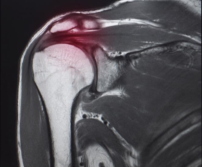
Generally, tendon injuries are caused due to trauma or gradual wear and tear from overuse or aging. Structurally, tendons—as well as their associated extracellular matrices—are comprised of nanostructured materials. Scientists have been focusing on tendon-bone healing using nanotechnology.

Image Credit: Yok_onepiece/Shutterstock.com
What is a Rotator Cuff Tear?
Rotator cuff tear (RCT) is a very common shoulder disease that causes pain in the shoulder and restricts activity. Scientists have reported that RCT is mostly prevalent among the older age groups and have estimated that around 20.7% of the world’s population suffers from RCT.
One of the most effective treatments of RCT is arthroscopic rotator cuff repair (RCR); however, a high rate of re-tear, ranging between 21% and 94%, has been recorded after the surgery.
Tendons are fibrous connective tissues that attach muscles to bones. Surgeons have indicated that poor tendon-bone healing in the rotator cuff is the main reason for the high rate of post-operative re-tears.
Generally, after repairing a rotator cuff, fibrovascular tissue is formed between the tendon and bone.
After the formation of the tissue, the bone grows into the fibrous interface, which slowly grows into the tendon. This tendon, subsequently, forms the continuous collagen fibers between tendon and bone.
However, surgeons have often observed problems with the development of tendons after surgery. After RCR, it is difficult for the bone to grow into the fibrovascular interface and tendon owing to the absence of blood vessels and bone loss at the tendon-bone junction.
As a substitute, the biomechanical inferior structure of scarred tissue is formed, which re-tears easily.
Thereby, researchers suggested that both osteogenesis (formation of new bones) and angiogenesis (formation of new blood vessels) are important for stimulating bone growth towards the tendon-bone junction. The phenomenon increases tendon-bone healing.
Role of Adenosine in Tendon–Bone Healing
Adenosine (Ade) is an organic compound present in human cells and plays the most important role in bone regeneration and maintaining bone homeostasis.
This compound is produced in ischemic, inflamed, or hypoxic environments, which can lower tissue injury. Ade also promotes tissue regeneration and repairs via a number of receptor-mediated mechanisms.
Ade acts on A2bR, as an autocrine/paracrine signaling molecule to elevate the osteogenic differentiation of stem cells.
Additionally, it is also a signaling molecule of tissue hypoxia and is responsible for the restoration of the blood supply in these tissues.
Previous studies have documented that Ade can upregulate the expression of vascular endothelial growth factor (VEGF) in cells, via hypoxia-inducible factor-1 (HIF-1α).
These studies have revealed the dual function of Ade, i.e., osteogenic and angiogenic activities. Hence, scientists believe that Ade can be quite beneficial in promoting tendon-bone healing.
Amorphous Calcium Phosphate Nanoparticles and Tendon–Bone Healing
Nanoparticles are materials of nano dimensions, i.e., less than 100 nm, with many unique properties. These materials are widely used in biomedical engineering to develop novel approaches for various areas of medicine such as oncology, nerve regeneration, tissue regeneration, radiology, etc.
Nanoparticles are used for various purposes such as the development of scaffolds, increasing mechanical strength and endurance, for their antimicrobial and anti-inflammatory properties, as carriers in gene therapy, and so on.
Researchers have suggested that owing to excellent biocompatibility and osteoinductive activity, calcium-phosphate-based biomaterials, e.g., calcium-phosphate matrix and hydroxyapatite, are commonly used for tendon-bone healing.
Recently, a group of scientists has successfully produced an organic amorphous calcium phosphate (ACP) mesoporous nanoparticle using the microwave-assisted hydrothermal method.
In this study, they used adenosine triphosphate (ATP) as the organic source for phosphorus.

Image Credit: Yurchanka Siarhei/Shutterstock.com
The main advantage of ACP mesoporous nanoparticles over the conventional calcium phosphate materials is their upgraded biocompatibility, high stability in aqueous solution, larger surface to area ratio, and an improved stable degradation curve.
Scientists revealed that during the synthesis of ACP nanoparticles, Ade is the by-product of ATP hydrolysis in an aqueous solution that possesses dual functions; namely, osteogenesis and angiogenesis.
Previously, ACP biomaterials had been used for tissue repairs, bioactive coating, and drug delivery. The unique use of ACP nanoparticles in tendon-bone healing is a new discovery.
In vitro and in vivo studies, using rat models with acute RCT, revealed that ACP nanoparticles could be effective in bringing about tendon-bone healing of the rotator cuff. This nanomaterial was rich in bioactive adenosine, which elevated osteogenesis and angiogenesis activities.
Radiological and histological tests showed that ACP nanoparticles increased the formation of bone and blood vessels at the tendon-bone junction.
Biomechanical evaluations have revealed that this nanoparticle has the ability to improve the biomechanical strength of the tendon-bone junction.
Thereby, this nanomaterial is identified as being the most appropriate biomaterial to promotes tendon-bone healing.
Other Nanoparticles Involved in Tendon Healing
Nanotechnology has improved extrinsic and intrinsic tendon healing post-surgery.
One of the techniques is the controlled release of mitomycin-C (chemotherapy agent) via hydrosol nanoparticles that reduces post-operative adhesions. Tendon tissue engineering uses nanocomposite scaffolds, e.g., modified silk nano scaffolds, for tendon healing.
Such scaffolding is better than allografts as it promotes an improved healing capacity with better mechanical stability.
Recent research has revealed that the transfection of miRNA by polylactic glycol acid (PLGA) nanoparticles can inhibit TGF-b1 expressions, which can help in the repair of a damaged tendon.
Another nanomaterial that has gained popularity in scaffold design is nano cellulose biomaterial.
Conclusion
This article has shown that several nanoparticles are associated with tendon healing.
Recent research has highlighted the potential of the newly developed ACP nanoparticles in tendon-bone healing. This is because they promote a dual biological function, i.e., osteogenesis and angiogenesis, which is essential for tendon-bone healing.
Scientists have proposed further research related to the repair conditions using ACP nanoparticles for a prolonged period.
References and Further Reading
Liao, H. et al. (2021) Amorphous calcium phosphate nanoparticles using adenosine triphosphate as an organic phosphorous source for promoting tendon–bone healing. Journal of Nanobiotechnology. 19, 270. https://doi.org/10.1186/s12951-021-01007-y
Qi, B. et al. (2021) Editorial: Applications of Nanobiotechnology in Pharmacology. Frontiers of Pharmacology. 10, pp.1451. https://doi.org/10.3389/fphar.2019.01451
Parchi, P. D. et al. (2016) Nanoparticles for Tendon Healing and Regeneration: Literature Review. Frontiers in Aging Neuroscience. 8, 202. https://doi.org/10.3389/fnagi.2016.00202
Yi, H. et al. (2016) Recent advances in nano scaffolds for bone repair. Bone Research. 4, pp. 16050. https://doi.org/10.1038/boneres.2016.50
Salata, O. (2004) Applications of nanoparticles in biology and medicine. Journal of Nanobiotechnology. 2(3). https://doi.org/10.1186/1477-3155-2-3
Disclaimer: The views expressed here are those of the author expressed in their private capacity and do not necessarily represent the views of AZoM.com Limited T/A AZoNetwork the owner and operator of this website. This disclaimer forms part of the Terms and conditions of use of this website.