Over the past two decades, spherical gold nanoparticles have been extensively employed in the research of photothermal cancer therapies.
However, these nanoparticles were not fully optimized due to the inherent limitations of spherical gold nanoparticles. Their peak absorptions, which reach a maximum of around 580 nm for particles with a 100 nm diameter, fall short of the 650 – 900 nm transmission window optimal for biological entities like skin, tissue, and hemoglobin. This discrepancy in absorption peaks limits their effectiveness in certain biological applications.
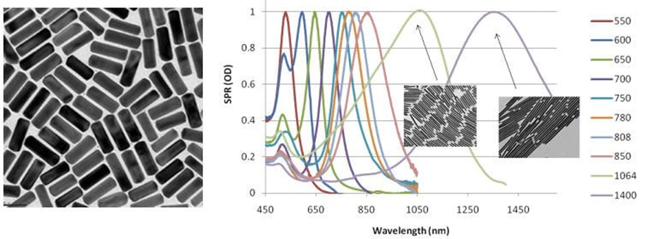
Gold nanorods behave in a similar manner to gold nanoparticles but are elongated to optimize their peak absorption and scattering characteristics, as illustrated in Figure 1. This elongation allows for the rate of nanorod absorption to be tuned from 550 nm to 1400 nm through different manufacturing processes, as shown on the right in Figure 1.
This tuning enables the gold nanorods to scatter at wavelengths across the visible and near-IR regions.
The surface plasmon resonance (SPR) value, indicating surface electromagnetic waves along the metal/solvent interface, is significantly influenced by materials adsorbed onto the metal surface or nanoparticles. Accurate absorption values for metal nanostructures can be achieved by measuring SPR values at various wavelengths, as depicted in Figure 1.

Figure 1. Left: TEM image of typical gold nanorods (10 nm × 40 nm) (Product No. 716820). Right: Gold nanorod peak absorption and scattering (extinction) can be tuned across the visible and near IR spectra shown on a plot of Surface Plasmon Resonance (SPR) to Wavelength. Image Credit: Merck
Gold nanorods are also finding application in enhancing in-vivo imaging through photoacoustics and highly efficient non-linear optics like four-wave mixing.
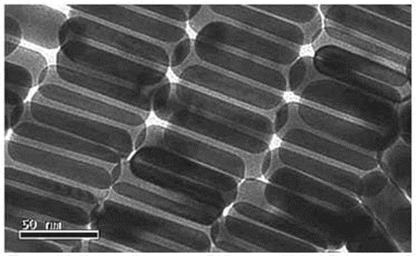
At the Centre for Micro-Photonics, Swinburne University of Technology, Victoria, Australia, researchers are actually using these nanorods as polarizers (Figure 2) and have successfully increased the storage capabilities of DVDs by over 2,000 times.

Figure 2. TEM image of gold nanorods used as polarizing materials. Image Credit: Merck
Materials
Source: Merck
| . |
. |
 |
Gold nanorods 10 nm diameter, λmax, 780 nm, dispersion in H2O |
 |
Gold nanorods 10 nm diameter, λmax, 808 nm, dispersion in H2O |
 |
Gold nanorods 10 nm diameter, λmax, 850 nm, dispersion in H2O |
 |
Gold nanorods carboxyl terminated, 10 nm diameter, λmax, 808 nm, dispersion in H2O |
 |
Gold nanorods palladium coated, 25 nm diameter, 100 μg/mL in H2O, dispersion |
 |
Gold nanorods 25 nm diameter, λmax, 600 nm, dispersion in H2O |
 |
Gold nanorods 25 nm diameter, λmax, 650 nm, dispersion in H2O |
 |
Gold nanorods 10 nm diameter, λmax, 980 nm, dispersion in H2O |
 |
Gold nanorods 10 nm diameter, λ: 1064 nm Amax, dispersion in H2O |
 |
Gold nanorods 10 nm diameter, Cy3 and methyl functionalized, powder |
 |
Gold nanorods 10 nm diameter, Cy3 and maleimide functionalized, powder |
 |
Gold nanorods 10 nm diameter, Cy3 and carboxyl functionalized, powder |
 |
Gold nanorods 10 nm diameter, Cy3 and biotin functionalized, powder |
 |
Gold nanorods 10 nm diameter, Cy3 and NHS functionalized, powder |
 |
Gold nanorods 10 nm diameter, Cy3 and azide functionalized, powder |
 |
Gold nanorods 10 nm diameter, Cy3 and streptavidin functionalized, powder |
 |
Gold nanorods 10 nm diameter, Cy3 and protein A functionalized, powder |
 |
Gold nanorods 10 nm diameter, Cy3 and amine functionalized, powder |
 |
Gold nanorods 25 nm diameter, FITC and maleimide functionalized, powder |
 |
Gold nanorods 25 nm diameter, FITC and amine functionalized, powder |
 |
Gold nanorods 25 nm diameter, FITC and protein A functionalized, powder |
 |
Gold nanorods 25 nm diameter, FITC and methyl functionalized, powder |
 |
Gold nanorods 25 nm diameter, FITC and streptavidin functionalized, powder |
 |
Gold nanorods 25 nm diameter, FITC and azide functionalized, powder |
 |
Gold nanorods 25 nm diameter, FITC and NHS functionalized, powder |
 |
Gold nanorods 25 nm diameter, FITC and biotin functionalized, powder |
 |
Gold nanorods 25 nm diameter, FITC and carboxyl functionalized, powder |
Citrate Capped
Due to the outstanding optical properties derived from surface plasmon resonance, gold nanorods are utilized in biomedical imaging, drug delivery, and photothermal treatment. Citrate-capped nanorods, in contrast to CTAB-capped nanorods, exhibit non-cytotoxicity and are particularly well-suited for biomedical applications.
Source: Merck
| . |
. |
 |
Gold nanorods 10 nm diameter, λmax, 780 nm, dispersion in H2O, citrate capped |
 |
Gold nanorods 10 nm diameter, λmax, 808 nm, dispersion in H2O, citrate capped |
 |
Gold nanorods 10 nm diameter, λmax, 980 nm, dispersion in H2O, citrate capped |
 |
Gold nanorods 10 nm diameter, λmax, 1064 nm, dispersion in H2O, citrate capped |
 |
Gold nanorods 25 nm diameter, λmax, 550 nm, dispersion in H2O, citrate capped |
 |
Gold nanorods 25 nm diameter, λmax, 650 nm, dispersion in H2O, citrate capped |