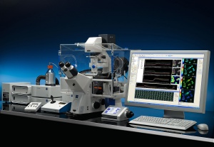
A new software module from Carl Zeiss enables real-time image capture and measurement of physiological parameters in living cells both during microscopic observation (online) and afterwards (offline). The AxioVision Physiology software captures the images and measurement data together with any changes in experimental parameters.
 The AxioVision Physiology software module from Carl Zeiss enables both image capture and the online as well as offline measurement of physiological parameters in living cells during microscopic observation.
The AxioVision Physiology Module is ideal for cell biologists, neurobiologists, physiologists and electrophysiologists. Applications include the determination of calcium concentrations or pH values through ratiometric calculation of fluorescence images after the addition of indicators, such as Fura-2 or Indo-1. The software also offers users an easy and reliable way to measure the change in fluorescence intensity over time of fluorescent proteins and for FRET analysis of protein interaction.
The Physiology Module integrates totally with Zeiss microscopes, enabling users to plan highly flexible experiments. In addition to the monochrome AxioCam cameras from Carl Zeiss, cameras from other manufacturers such as Hamamatsu or Roper are supported. Two synchronously controlled AxioCam cameras are also supported when combined with the Carl Zeiss Dual Camera Module. Parameters, such as image capture frequency, are easily changed during the course of the experiment and the software supports external light sources like the Zeiss Colibri or Sutter DG4 and other external components. The direct streaming of the camera data on the hard drive means that the technically possible image capture rate of the cameras can be achieved without limiting the experiment duration.
Combined with the Axio Observer.A1 manual microscope, the AxioVision Physiology Module makes a perfect entry-level physiological measurement station. The module physiology software may also be combined with the Cell Observer microscope system, based on the motorized Axio Observer.Z1. Maximum flexibility and performance is available by combining the new software with the Axio Examiner fixed stage microscope.