Carl Zeiss has developed a unique series of solutions addressing the different methods for brain mapping and soft tissue imaging. "Scientists are right now attacking one of the last secrets of mankind: imaging and reconstruction of the brain", Dr. Dirk Stenkamp, Member of the Board at Carl Zeiss SMT explains. "We specifically enable the acquisition and analysis of cell images at ultra-high resolution. For that purpose we have developed an extensive range of solutions, based on the sophisticated use of advanced electron and ion-beam microscopes", Stenkamp adds.
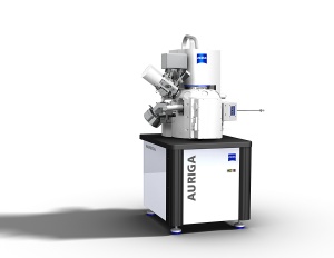
 The new AURIGA CrossBeam workstation from Carl Zeiss offers unique imaging, advanced analytics and precise processing.
At center stage, with a combination of a special detector system and large framestore, the SIGMA™ FE-SEM is enabling extremely fast imaging of huge areas of thin sections. "With this system, which is currently being evaluated by several leading research institutes worldwide, throughput is increased by a factor of 100", application product specialist Dr. Stewart Bean from Carl Zeiss SMT´s Cambridge facilities says.
Extremely thin sections can be investigated by simultaneous milling and imaging using ZEISS CrossBeam® workstations like the NVision 40 or the new AURIGA. This leading-edge FIB/SEM instrument provides high resolution and now also features the possibility of investigating nonconductive samples using a unique local charge compensation method. NVision CrossBeam systems are already successfully in the field for soft tissue analysis at various institutes, including EPFL in Lausanne (Switzerland) and many others.
The ORION® Helium-ion microscope offers totally new imaging possibilities. "Biological samples especially profit from the unprecedented depth of focus and the high contrast, that are inherent characteristics of imaging with helium ions," Mohan Ananth, Product Manager and application specialist at the US headquarters of Carl Zeiss SMT points out.
Whenever resolution below one nanometer is required, the LIBRA® 120 PLUS energy filtering TEM from Carl Zeiss SMT provides the highest contrast imaging of soft tissue. LIBRA 120 PLUS especially matches the extreme demands in revealing structural and 3-D information of beam sensitive or frozen hydrated specimens at the nano scale. With the LIBRA 200 FEG energy filtering TEM, resolution in the Angström and even sub-Angström range can be provided.
"We are in dialogue with researchers all over the world in order to meet their demanding requirements – now and in the future and as a part of our company mission. We are proud to be making a contribution to research work that will change our world", Stenkamp explains.