Scientists who discovered an innovative three-dimensional microscope method are extending this technology to be suitable for a range of applications in biological research, medicine, and production of novel electronic equipment.
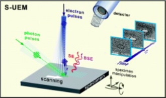
 Scientists are describing development of a revolutionary instrument called a 4-D scanning ultrafast electron microscope
They published reports on a new technology known as four-dimensional scanning ultra-fast electron microscopy and another related method that appeared in two separate papers in the Journal of the American Chemical Society.
Ahmed H. Zewail, a Nobel Laureate in Chemistry with his coworkers converted high-resolution three dimensional images of objects at the nanoscale level to four dimensions when they found a method of integrating time into conventional electron microscopy observations.
Their laser-based technology enabled researchers to see three-dimensional formations including carbon nanotubes in the shape of a ring which twisted on heating, over a minute time range of femtoseconds. The researchers could not get much three-dimensional data using that technique because it was not capable of capturing the natural motion of objects and the objects seemed as if they were stationary.
The scientists explain how four-dimensional scanning transmission ultrafast electron microscopy overcomes that challenge, and provided detailed information about the material’s innermost structures.
The reports demonstrate how the method will prove useful to examine atomic-scale dynamics on the surfaces of metal, and observe the vibrations of a gold nanoparticle and an individual silver nanowire. The researchers explain that new methods can be used for a wide range of applications in single-particle biological imaging and in materials science.