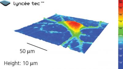
The need for powerful tools for understanding the fine inner functionings of neurons is critical. By using a technology from materials science as the basis, a research team comprising advanced imaging experts, psychiatrists, and neurobiologists from Switzerland's CHUV and EPLF has identified a new way to use digital holographic microscopy (DHM) for observing in real-time the neuronal activity at three-dimensions with a resolution of up to 50 times greater than existing methods.
 This is a 3-D image of living neuron taken by DHM technology. Credit: Lyncée Tec
The understanding of DHM is made simple with this illustration. When the sea waves hit a large rock and flow out the other side, they carry with them information of the rock's shape. By comparing this information with the waves that did not contact the rock, the rock’s image can be designed. DHM applies the same concept using a laser beam which is made to focus a single wavelength on a neuron sample. The distorted wave was collected on the other side and then compared with a normal beam. Using an algorithm, a computer numerically designs the three-dimensional image of the sample. Besides, the laser beam passes through the transparent neuron cells and carries with it vital information of its internal composition.
Generally, DHM was used to identify miniature defects present in materials. Now, the research team has ascertained to apply DHM in neurobiological purposes. In this research, the team stimulated an electric charge in the neurons’ culture making use of glutamate which is regarded as the brain’s key neurotransmitter. Water is being transported by this electric charge into the neurons which alters their optical characteristics in a manner that only DHM can discover them. Hence, this approach allows real-time observation of several hundreds of neurons’ electrical properties at the same time preventing the damage caused by the electrodes, capable of recording few number of neurons’ activity simultaneously.
>
3-D Dynamic Images of Neurons @EPFL
DHM can be utilized in high content screening, which is the screening of a large number of newly created pharmacological molecules, without employing electrodes and dyes. This technique can be utilized for testing newly created drugs against neurodegenerative diseases including Parkinson's and Alzheimer's as it is capable of testing more number of new molecules rapidly.