A research team at the University of Illinois has devised a novel imaging technique called spatial light interference microscopy (SLIM) that uses two light beams to quantify cell mass.
The SLIM technique, a combination of holography and phase-contrast microscopy, could resolve the issue of whether cells develop exponentially or at a constant rate. This highly sensitive technique measures cell mass with femtogram precision. It can quantify a single cell growth as well as mass transport inside the cell.
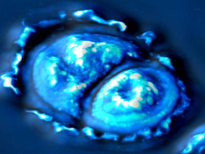
 SLIM-imaged cell. Credit: Quantitative Light Imaging Laboratory
The fully non-invasive method, SLIM technique allows the researchers to investigate cells when they do their usual functions. It utilizes white light and can be coupled with additional conventional microscopy technologies, such as fluorescence, to study cell growth.
The high sensitivity of the SLIM technique enables the researchers to observe cell growth at various cell cycle phases. They observed that mammalian cells grow exponentially only during the cell cycle’s G2 phase, before the cell divides and after the DNA reproduces. This finding is important not only for fundamental biology but also for tissue engineering, drug development and diagnostics.
The researchers plan to utilize the new information of cell growth to various disease models. They intend to utilize the SLIM technique to measure the growth rate between cancer cells and normal cells as well as the impact of curing methods on the growth rate.
Gabriel Popescu, who led the development of the SLIM technique, is launching SLIM as a common resource in the University of Illinois campus, expecting to control its versatility for fundamental and clinical research in various fields.