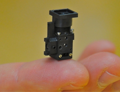
Scientists at the Stanford University have designed a fingertip sized portable microscope weighing 1.9 g, which is designed to view fluorescent markers or dyes that are broadly utilized by biological and medical scientists to study the brain of mice.
 Fingertip-size portable microscope
Absence of moving parts, which need repositioning for better resolution of the scope, and protection against dust make the three-quarters tall microscope ideal for utilization outside the laboratory. The microscopic image can then be viewed on a computer or laptop. The components of the device were actually designed for mobile phones and other consumer electronics and can be bulk-produced at low costs. The portable microscope has better resolution and optical sensitivity compared to fiberoptic or traditional desktop microscopes.
The research team comprising research group of Mark Schnitzer and Abbas El Gamal had originally planned to design a novel custom chip but it has changed its plan to develop a system instead. Prior to actually assembling the components, the team designed a virtual way to test probable combination of components in a computer simulation. The final product functioned at the desired level of performance.
Schnitzer's group has been investigating a specific area in the mouse brain that is responsible for the co-ordination of the fore and hind limbs during movement. It used an ultra-thin set of wires instead of fiberoptics in order to connect the computer and the microscope. This method allows the mouse to move more freely. With the combination of better resolution, broader field of view and improved mobility, the research team observed an unprecedented pattern in which neurons were fired concurrently in bands of a maximum of 30 neurons when the mice moved. Schnitzer stated that such a pattern occurs only during movement of the animals.
The scientists have utilized the novel microscope to determine tuberculosis in cells developed in the lab. They have also utilized the device by combining four of them in an array to count individual cells grown in the lab and attained a precision equivalent to that of a standard full-size cell counting device. They have also illustrated the device’s ability to screen certain genetic mutations. El Gamal stated that the device’s image resolution can be enhanced to 1 µm from the current value of 2.5 µm by utilizing advanced cell phone imagers.