A research team at Purdue University is devising a technique using an atomic force microscope to determine the mechanical characteristics of live cells, paving the way to efficiently analyze biological processes and diagnose human diseases.
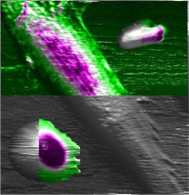
 This artist's conception depicts the use of an atomic force microscope to study the mechanical properties of cells, an innovation that might result in a new way to diagnose disease and study biological processes. Here, three types of cells are studied using the instrument: a rat fibroblast is the long slender cell in the center, an E coli bacterium is at the top right and a human red blood cell is at the lower left. The colored portions show the benefit of the new technique, representing the mechanical properties of the cells, whereas the gray portions represent what was possible using a conventional approach. (Credit: Purdue University image/Alexander Cartagena)
The technique can be utilized to investigate the adhesion of cells to tissues, which is important for most biological processes and diseases, the evolution of cancer cells during metastasis, the capability of cells to travel and modify its shape, the response of cells to mechanical stimuli required for stimulation of key protein synthesis.
An atomic force microscope is suitable to study the mechanical properties of the smallest cellular structures, as it can view tiniest objects that cannot be imaged utilizing light microscopes. The innovative Purdue University technology is around five folds quicker than other techniques using atomic force microscopes. Its ultra-speed capability allows real-time observation of biological processes and living cells. With this method, a ‘mechanobiology-based’ assay can be developed to support the typical biochemical assays.
Arvind Raman, one of the members of the research team, stated that the innovative technique enables mapping to determine the mechanical properties of various cell parts, whether they are squishy or rigid or soft. The novel technique offers high-resolution mapping at higher speed when compared to traditional methods, mainly due to advancements in signal processing, he added.
Raman further said that the research team used the innovative technique to analyze three different types of cells, including rat fibroblasts, human red blood cells and bacteria, in order to prove its capability in research and medicine.