Scientists at the University of Illinois have published the results of their studies to evaluate microscopy techniques employed to analyze the structure and shape of pollen grains.
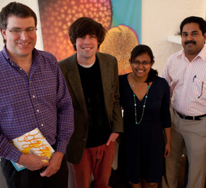
 University of Illinois researchers (from left) Glenn Fried, Luke Mander, Surangi W. Punyasena and Mayandi Sivaguru
Classification or taxonomy of flora is largely dependent on the study of pollen morphology. Conventional methods in use to study the image of pollen grains are reflected and transmitted light techniques. But they have their limitations and do not provide an image good enough for conclusive analysis. Reflected light method for instance provides a good image of the pollen shape but fails to provide a fine surface texture resolution. Transmitted light technique does not provide a good image of the shape but can provide fine texture resolution. The research team led by Mayandi Sivaguru and Surangi Punyasena at the Institute of Genomic Biology used three samples of pollen grains of differing texture and grain size to compare the imaging results from the various microscopy methods available. They found that the effective technique was a combination of both reflective and transmitted light imaging.
Luke Mander, co-author of the research paper, stated that the study would help resolve limitations in flora classification. A typical challenge is the classification of grass species which number at 11,000. The pollen produced by all the species is similar and makes it difficult to determine the diversification history of grass from fossilized pollen grains. The super-resolution structured illumination (SR-SIM) technique of imaging employed by them has the capability to image structural features smaller than 200 nm. This will help to understand the composition and evolution of the grasslands. The researchers are confident that this capability to image morphology below the light diffraction limit will pave the way for SR-SIM to replace electron microscopy for future pollen analysis.