Oct 8 2014
Cal Poly Pomona has received a $203,000 Major Research Instrumentation Program award from the National Science Foundation to purchase a scanning electron microscope (SEM).
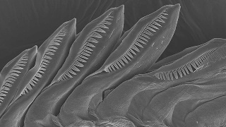
 An image of a sea slug's teeth taken through a scanning electron microscope. Cal Poly Pomona has received a National Science Foundation grant to purchase one of the microscopes.
The microscope will be housed in an engineering lab, but will be accessible to other departments, industry-sponsored projects and community outreach programs. Several faculty members have plans to utilize the microscope in research projects, and it also will be incorporated into classroom curriculum for majors in science and engineering.
The microscope uses electrons, instead of visual light as in a traditional optical microscope. SEM can be used on many types of materials, including, metals, ceramics, polymers or biological samples.
In the SEM a beam of electrons is sent onto a material surface, creating a number of electron interactions with the sample. Through these interactions, emergent electrons can be gathered to create an image. Different information can be obtained based on what type of electrons are captured, such as secondary or backscattered electrons.
The images created provide detailed information about the components of the materials, including chemical composition on a microscopic level and topographic data. The SEM will give new insights that a traditional microscope cannot provide.
“A SEM provides multifaceted imaging abilities,” says Vilupanur Ravi, chair of the chemical and materials engineering department and principal investigator on the project. “It gives us a new level of capability and allows us to do cutting-edge research.”
Angel Valdes, associate professor of biology and a co-principal investigator, drives to the Natural History Museum in Los Angeles to use its SEM for his research on sea slugs. He looks forward to having one on campus.
“The SEM is a powerful tool to identify problematic species and understand the evolution of morphologic traits in sea slugs and other organisms. I include SEM photographs in almost all my papers,” he says. “The SEM is a critical teaching tool that I use to train my graduate and undergraduate students in the anatomical characteristics of sea slugs.”
Once the SEM is on campus, the science and engineering departments will enhance their curriculum with new classes.