May 12 2015
Researchers from the Zelinsky Institute of Organic Chemistry of Russian Academy of Sciences have developed an easy to use procedure for visualizing defects on graphene layers.
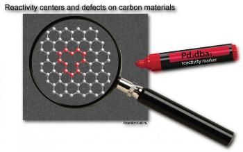
 Location of defects is important to estimate the quality of carbon materials and to predict physical and chemical properties of graphene systems. Credit:Ananikov Lab
This study was part of an international project where they mapped the surface of carbon materials using a suitable contrast agent.
On large carbon surface areas, tomography has the ability to show organized defect patterns. Tomography is a microscopic imaging procedure that can capture different types of defects within a short duration.
2D materials such as graphene are considered to become valuable compounds of the century. Graphene demonstrates remarkable electrical and thermal properties and is very strong though it is very thin.
Graphene is considered to hold significant promise for creating new smart materials, efficient light conversion, photocatalysis and also for biomedical applications.
However, a deterrent has been the requirement of perfect graphene having a specific number of defects. It is difficult to prepare graphene surfaces that are free of defects, as well as the size and shapes of the defects.
Furthermore, the defects cannot be detected easily due to fluctuations and dynamic behavior. It is cumbersome, time consuming process when large graphene sheets have to be scanned for locating defects.
Ananikov and co-workers had conducted joint research which showed that soluble palladium (Pd) complex had the ability to selectively attach itself to defective areas on the carbon material’s surface. Soluble palladium complex is a contrast agent.
Nanoparticles were formed when Pd attached to the defects, and these can be detected with a normal electron microscope. When the carbon center is more reactive, the contrast agent binds more strongly. Utilizing a handy nanoscale imaging procedure, the defect sites and reactivity centers were mapped in 3D space with high contrast and resolution.
This procedure identified carbon defects based on their varying chemical reactivity and difference in morphology. This innovative process enables chemical reactivity visualization with spatial resolution.
Unique insight of the graphene layer’s reactivity is obtained by using "Pd markers" to map the carbon reactivity. This study showed that in 1μm2 of carbon material surface, over 2000 reactive centers could be located.
The carbon material’s spatial complexity at the nanoscale has been reveled by this study. Significant amount of gradients and variations over the surface area were observed by mapping the surface defect density.
Tomography has been successfully used for medical diagnostic applications for many years. Contrast agents have been used to obtain better and more accurate observations. This study demonstrates the possibility of using tomography applications for conducting atomic scale diagnostics of materials.
The research team has published this study as an article “Spatial imaging of carbon reactivity centers in Pd/C catalytic systems” in the Chemical Science journal of the Royal Society of Chemistry.