May 23 2016
An international group of scientists from Russia and France, in collaboration with researchers from the Lomonosov Moscow State University, have successfully produced ultrapure silicon nanoparticles, which possess the characteristic of photoluminescence, that is, secondary light emission following photoexcitation.
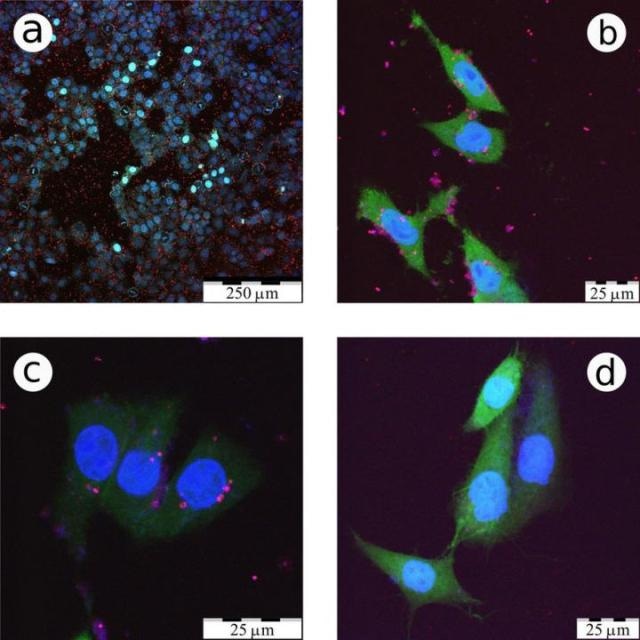
 These are confocal fluorescence microscopy images of CF2Th cancer cells incubated with LA-Si NPs. (Credit: Victor Timoshenko/Scientific Reports)
These particles can easily enter cancer cells, allowing them to be used as luminescent markers in the early diagnoses and treatments of cancer.
The results of the study have been reported in the Scientific Reports journal.
Many laboratories around the globe are actively exploring different ways to develop these types of nanoparticles, but according to Victor Timoshenko, one of the study co-authors, professor of the Physics Department of the Lomonosov Moscow State University, the quality of the particles was poor, because chemical reactions in acid solutions were used for synthesizing them.
The obtained particles were not sufficiently pure. By-products of the chemical reactions made them toxic. Furthermore, these nanoparticles had a form, which was far from a sphere, and it does not contribute to the appearance of the photoluminescence. These two drawbacks severely restricted their applications.
Victor Timoshenko, Professor of Physics, Lomonosov Moscow State University
To resolve these drawbacks, the team applied a new approach that involved the use of laser ablation, with no previous positive outcomes. In laser ablation, atoms are ejected from the target with a beam of laser, so that the atoms that were torn would form a nanocrystal. The issue here was that the torn atoms in this situation do not integrate with particles, but rather combine to certain random layers, and they also do not glow after acquiring the nanoparticles. Here, either the nanoparticles cool down too rapidly or they were very large, and lacked the time to form nanocrystals of high-quality. To put it simply, it was essential to warm these particles so that crystallization occurs at least for a brief period of time.
For that purpose, we decided to use high-intensity, short laser pulses. They not only ejected the silicon atoms from the target, but additionally ionized them. The emitted electrons led to the ionization of helium atoms, in which atmosphere it all was happening. In a very short time of nanoseconds something of a microwave kind appeared, laser plasma formed the conditions that allowed the atoms to sinter into spherical nano-crystals. These beads falling onto the surface aggregated as a fluffy layer, which subsequently could be readily dispersed in water.
Victor Timoshenko, Professor of Physics, Lomonosov Moscow State University
These particles were round in shape and had the right size, measuring 2 to 4 nm in diameter, which offered efficient photoluminescence where individual falling photons are balanced with a single ejected one.
Contrary to the nanoparticles acquired by a chemical etching process, they lacked lethal additives, and most significantly, as shown by biological experiments, the nanoparticles can easily enter into the cells. Such nanospheres penetrate into the cancerous cells much more readily than that of the healthy ones.
This can be attributed to the fact that the cancerous cells are always ready to split and tend to absorb the surrounding environment to produce daughter cells. Victor Timoshenko informed that when compared to the healthy cells, cancer cells absorb nanoparticles 20 to 30% more efficiently based on the type of cells, and this can lay the framework for the early-stage cancer diagnosis.
Our main achievement was that we produced such nanoparticles and established that they easily penetrate into cancer cells. The problem of the diagnostic is a separate task, which is solved simultaneously by biologists, with our participation. You can, for example, replace the analysis of biopsy, a fairly long and not too reliable "yes-no" test, in which the cancer cells in the body are detected by the fact whether a nanoparticle penetrated a tissue sample, or it did not. There are also non-invasive diagnostic methods. The photoluminescent light emitted from the nanoparticles in this case is difficult to use, but they can be activated by other means, for example, ultrasound or radio frequency electromagnetic waves.
Victor Timoshenko, Professor of Physics, Lomonosov Moscow State University
The acquired nanoparticles are fully non-toxic and are excreted easily. Another important aspect is that these nanoparticles bind to certain substance or class of biomolecules like antibodies to their surface, making it easy to target them to penetrate into the cancer cells and thus improving the efficiency of diagnosis.
Victor Timoshenko added that going forward, the acquired nanoparticles will be attached with drugs, which would both help detect cancer and perform a localized radiotherapy or chemotherapy at the cellular level.