Every year, the WITec Paper Award competition recognizes three exceptional peer-reviewed publications that feature results acquired with a WITec microscope. A record number of 113 publications was submitted this year, in a clear demonstration of the power of Raman imaging for diverse fields of application such as cancer research, electrochemistry, semiconductor research, geology and microplastics research, to name only a few. WITec thanks all participants for their outstanding contributions from all over the world. The Paper Awards 2020 go to researchers from Japan, Poland and Austria and acknowledge impressive studies and methodologies from the fields of electrochemistry, biomedicine and polymer science, respectively.
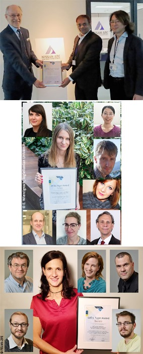
 The winners of the WITec Paper Award 2020. Top: Gold winners Ankur Baliyan (middle) and Hideto Imai (right) together with WITec K.K. representative director Michael Verst (left). Middle: Silver winner Ewelina Wiercigroch (middle) and her co-authors (clockwise, beginning top left): Elzbieta Stepula, Yuying Zhang, Lukasz Mateuszuk, Kamilla Malek, Sebastian Schlücker, Malgorzata Baranska and Stefan Chlopicki. (© E. Wiercigroch) Bottom: Bronze winner Ruth Schmidt (middle) and her co-authors from left to right: Armin Zankel, Claudia Mayrhofer and Hartmuth Schröttner (top row); Manfred Nachtnebel and Harald Fitzek (bottom row). (© FELMI-ZFE)
- GOLD: Ankur Baliyan and Hideto Imai (2019) Machine Learning based Analytical Framework for Automatic Hyperspectral Raman Analysis of Lithium-ion Battery Electrodes. Scientific Reports 9: 18241. doi.org/10.1038/s41598-019-54770-2
- SILVER: Ewelina Wiercigroch, Elzbieta Stepula, Lukasz Mateuszuk, Yuying Zhang, Malgorzata Baranska, Stefan Chlopicki, Sebastian Schlücker and Kamilla Malek (2019) ImmunoSERS Microscopy for the Detection of Smooth Muscle Cells in Atherosclerotic Plaques. Biosensors and Bioelectronics 133: 79-85. doi.org/10.1016/j.bios.2019.02.068
- BRONZE: Ruth Schmidt, Harald Fitzek, Manfred Nachtnebel, Claudia Mayrhofer, Hartmuth Schröttner and Armin Zankel (2019) The Combination of Electron Microscopy, Raman Microscopy and Energy Dispersive X-Ray Spectroscopy for the Investigation of Polymeric Materials. Macromolecular Symposia 384: 1800237. doi.org/10.1002/masy.201800237
For a list of all previous Paper Award winners, please visit www.witec.de/paper-award.
The Paper Award GOLD: Automated Quality Control of Lithium-Ion Batteries
Lithium-ion batteries (LIBs) provide the power for most electric devices that we use every day, such as cell phones, tablets and laptops. Their development was honored with the Nobel Prize in Chemistry last year. Automated real-time quality control of LIB materials is necessary for industrial research and production. Ankur Baliyan and Hideto Imai from Nissan Arc. (Yokosuka, Japan) win the Gold Paper Award 2020 for their machine learning-based approach to analyzing Raman data of LIBs. Raman images of LIB cathodes can visualize the spatial distribution of the active cathode material (lithium nickel manganese cobalt oxide, abbreviated as LiMO2) and the surrounding carbon matrix. In order to automate and accelerate the process of identifying the spectral signatures in Raman datasets, the authors developed a machine learning-based analytical framework. It starts by automatically pre-processing the Raman data to remove the baseline and cosmic rays. Next, algorithms determine the number of components, extract the corresponding spectral signatures, and identify them. The spectra are finally used to train a neural network, which can then automatically analyze Raman data from the same or a different LIB sample. The authors demonstrated that data analysis by the trained neural network yielded results consistent with results from an experienced user. However, the algorithm found two minor signatures in addition to the main components of carbon and LiMO2 that corresponded to a residual background signal and one of the main components exhibiting increased fluorescence signals. The presented approach requires very little user input and is thus suitable for real-time quality control using Raman data from lithium-ion batteries and other applications.
The Paper Award SILVER: Characterizing Atherosclerotic Plaques with iSERS Microscopy
“Atherosclerosis is one of the major causes of death worldwide. Understanding the mechanism of its formation still remains a great challenge in medicine. Powerful techniques for monitoring the composition and stability of atherosclerotic plaques are thus needed,” says Ewelina Wiercigroch from Jagiellonian University (Krakow, Poland), winner of the Silver Paper Award 2020. Atherosclerotic plaques form at arterial walls and narrow the blood vessels. Monitoring the stability of the plaques is of clinical relevance because their rupture can result in a stroke or heart attack. As smooth muscle cells (SMCs) play a key role in stabilizing the plaques, their presence can serve as a marker for plaque stability. Ewelina Wiercigroch, Elzbieta Stepula, Lukasz Mateuszuk, Yuying Zhang, Malgorzata Baranska, Stefan Chlopicki, Sebastian Schlücker and Kamilla Malek from Jagiellonian University and the University of Duisburg-Essen (Germany) demonstrated the suitability of immuno surface-enhanced Raman scattering (iSERS) microscopy for staining SMCs in atherosclerotic plaques. SERS labels were conjugated either with a primary antibody directed against α-actin of SMCs (direct iSERS) or with an appropriate secondary antibody (indirect iSERS). The iSERS images of mouse artery sections visualized regions containing SMCs and cluster analysis allowed the quantification of the percentage of SMCs located in the plaques. Results from iSERS staining agreed qualitatively and quantitatively with those from immunofluorescence (IF) staining. IF is the current gold standard in visualizing atherosclerotic constituents, but iSERS offers some advantages, such as higher photostability. The study thus establishes iSERS as a promising technique for visualizing and quantifying SMCs in atherosclerotic plaques.
The Paper Award BRONZE: Correlative Raman Imaging of Polymeric Materials
Ruth Schmidt from Graz University of Technology (Graz, Austria) receives the Bronze Paper Award 2020, together with her colleagues Harald Fitzek, Manfred Nachtnebel, Claudia Mayrhofer, Hartmuth Schröttner and Armin Zankel. The group demonstrated the potential of correlative Raman Imaging and Scanning Electron (RISE) microscopy and Energy Dispersive X-Ray Spectroscopy (EDXS) for investigating polymers. Polymeric materials are popular in many applications due to their wide variety of useful properties, such as high elasticity or toughness. For characterizing their properties, the advantages of three imaging techniques were combined. Scanning Electron Microscopy (SEM) acquired high-resolution structural information, while Raman imaging revealed the chemical composition and was complemented by elemental information from EDXS. The publication provides a detailed methodology chapter which describes different sample preparation approaches and imaging modes. For example, it explains strategies for SEM imaging without coating the sample, which would hinder subsequent Raman imaging. Three polymer specimens were investigated with RISE and EDXS, yielding complementary information from the same sample region. Coarse and fine structures of the samples were correlated with chemical properties and the layer structure of packaging materials was visualized. Particulate additives in a polymer matrix were identified and their size distribution was investigated. The authors stressed that combined SEM, Raman imaging and EDXS offers great possibilities for analyzing polymeric materials.
The Competition Continues: WITec Paper Award 2021
Scientists from all fields of application are invited to participate in the Paper Award 2021 contest (www.witec.de/paper-award). Articles published in 2020 in a peer-reviewed journal that feature data obtained with a WITec microscope can be submitted to [email protected] until January 31st, 2021. WITec is again looking forward to receiving many new and exceptional publications.