While the novel nanomaterial graphene is being utilized for various applications, its effect on actin filament dynamics has remained uncharted territory. Innovative research published in The International Journal of Molecular Sciences has uncovered this area of science to observe and possibly develop actin assembly kinetics.

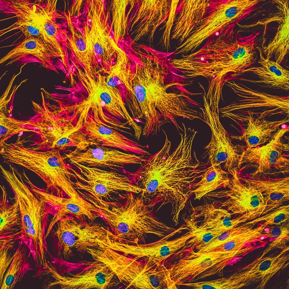
Study: Graphene Enhances Actin Filament Assembly Kinetics and Modulates NIH-3T3 Fibroblast Cell Spreading. Image Credit: Vshivkova/Shutterstock.com
Significance of Actin Research
Actin is considered to be a critical cytoskeletal protein as well as significant within the filaments of muscle fibrils. This protein can promote the reorganization of cellular structures through enabling processes such as cell morphogenesis, migration and differentiation.

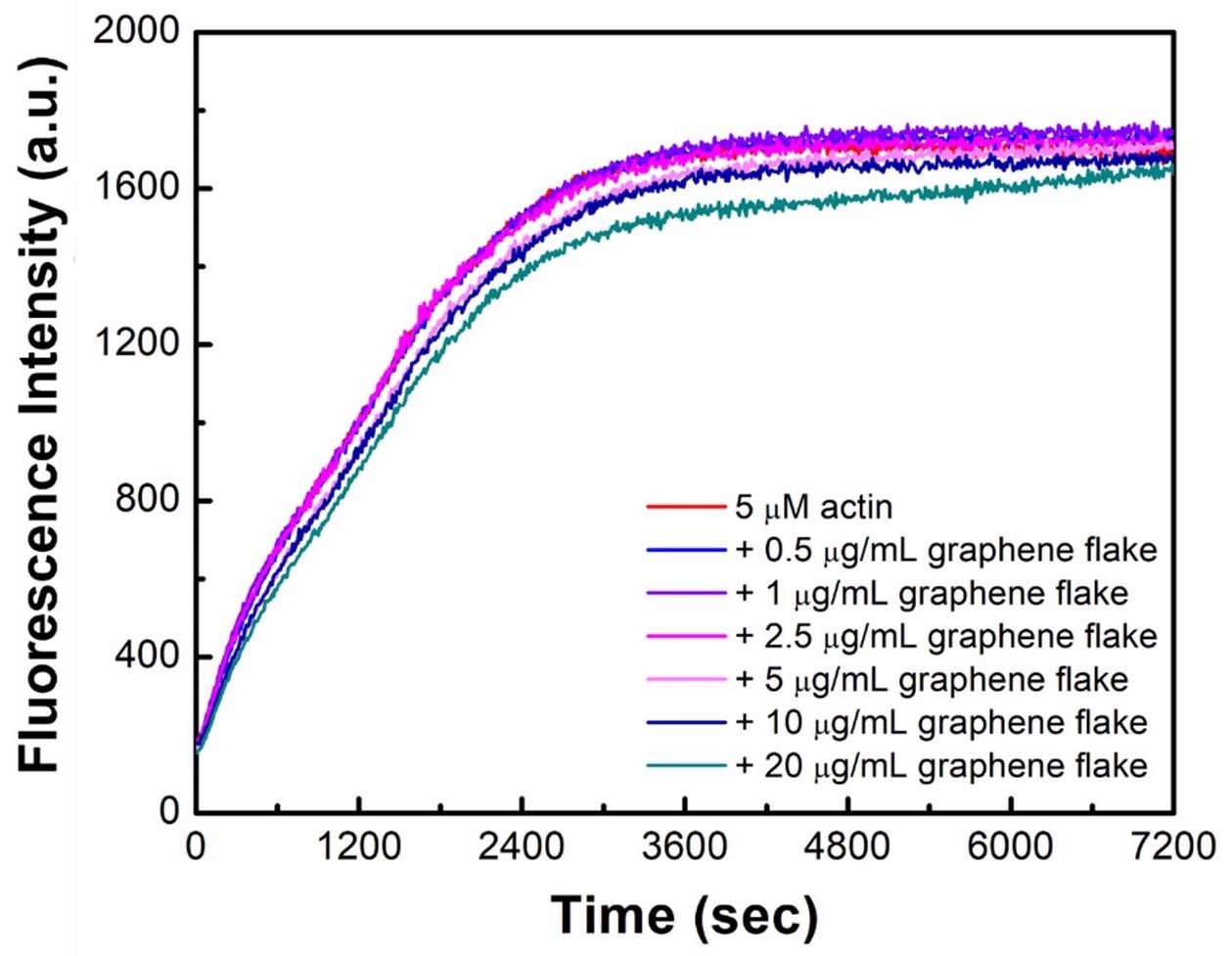
Graphene flakes do not hamper bulk actin polymerization. Actin (5 µM, 20% pyrene-labeled) polymerization in the presence of graphene flakes at varying concentrations (0.5–20 µg/mL) was monitored. Data are a representative from triplicated trials. © Park. J., Kravchuk. P., Krishnaprasad. A., Roy. T., and Kang. E.H. (2022)
Recent research has explored the use of nanomaterials on actin and found that exposure to nanomaterials, and carbon-based materials especially, including carbon nanotubes and graphene, has the ability to modulate actin polymerization. This research has given rise to other exciting research investigations, including how graphene can be used to enhance actin filament assembly.
Advancing Biomedical Applications Through Graphene
Graphene, a nanomaterial allotrope of carbon, consists of a single layer of atoms in a two-dimensional honeycomb lattice structure; it has remarkable physiochemical properties which enable the advancement of several fields involving chemical, electric, thermal and mechanical applications. These properties allow graphene to be desirable for industries including technology to medicine, for advancing electronics as well as use as drug delivery systems and cancer therapy.
Further research into how graphene can be utilized for cellular applications could enable further advancement in medicine; a key starting point is understanding how graphene can affect actin filament assembly dynamics.
Existing literature has shown the impact of graphene on actin rearrangement in naïve macrophages through increasing cytokine and chemokine production and reducing cell adhesion. Additionally, other research has suggested the accumulation of graphene nanoflakes on monkey kidney cells can cause reactive oxygen species production and then actin filament rearrangement.
Further reports have also touched on graphene nanoflakes leading to electron transfer disruption in mitochondria and reduced energy source from ATP, resulting in faulty actin filament assembly within breast cancer cells.
The implications of utilizing graphene to reorganize actin filaments for impaired cellular applications can be exploited for use against cancer – an exciting area of medicine that could benefit cancer therapy.

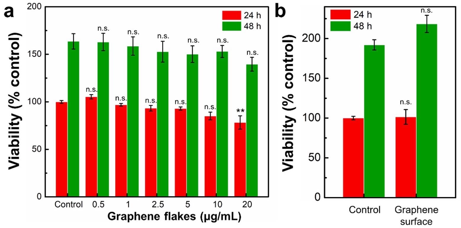
Effects of graphene flakes on viability of NIH-3T3 cells. The viability of NIH-3T3 cells was was determined after 24 h and 48 h exposure to various concentrations of (a) graphene flakes (0.5–20 µg/mL) or (b) on a graphene surface using a WST-1 assay. The results are expressed as the mean ± standard deviation (S.D.) of three independent experiments. n.s., not significant; **, p < 0.01. © Park. J., Kravchuk. P., Krishnaprasad. A., Roy. T., and Kang. E.H. (2022)
Innovative Research
The research team investigated the effect of graphene on the modulation of actin filament assembly kinetics, with a hypothesis that the non-covalent interaction between actin and graphene could potentially have a direct impact on actin filament assembly.
They utilized methods such as total internal reflection fluorescence (TIRF) microscopy imaging and bulk pyrene fluorescence assay and investigated cytotoxicity and morphology by using mouse embryo fibroblast NIH-3T3 cells seeded on a graphene surface.
The team found that both graphene flakes and a graphene surface significantly enhance the rates of actin filament elongation with cell culture experimentations illustrating a lack of cytotoxicity.
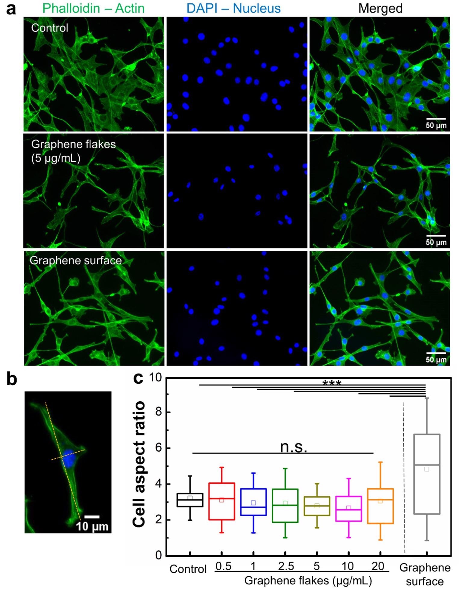
Additionally, the graphene surface may have enabled changes within the mouse embryo fibroblast cells, affecting cell spreading and stretched cell morphology. Interestingly, this suggests that exposure to graphene and subsequent interaction may directly impact molecular remodeling of the actin cytoskeleton with further impact on cell motility and physiology modulation.
Implications for Future Therapies
Actin holds a very significant role in cellular applications, with significant functions for its many forms, from actin binding proteins involved in the formation of cross-links to actin isoforms such as ACTA1, a gene encoding the skeletal muscle α-actin, critical for muscle contraction. ACTA1 gene expression is also altered in many cancer types, such as being downregulated in head and neck squamous cell carcinomas, a key association to tumorigenesis. Additionally, it can also be used as a biomarker for chemoresistance in basal-like breast cancer.

Effects of graphene flakes and graphene surface on NIH-3T3 cell morphology and spreading. (a) NIH-3T3 cells were incubated for 24 h on either a poly-L-lysine coated coverslip (top), on a poly-L-lysine coated coverslip and treated with graphene flakes (5 _g/mL) (middle), or on a pristine graphene monolayer (bottom). Then, cells were stained with Acti-stain 488 phalloidin (actin, green) and DAPI (nucleus, blue). (b) Representative confocal microscopy image of a cell used to measure the cell aspect ratio (major/minor axis). (c) Quantified cell aspect ratio of NIH-3T3 cells incubated with graphene flakes or on a graphene surface. N = 20–73 across three independent experiments. n.s., not significant; ***, p < 0.001. © Park. J., Kravchuk. P., Krishnaprasad. A., Roy. T., and Kang. E.H. (2022)
The potential applications of actin remodeling could be utilized for therapies that target actin expression in cancer, enhanced through the use of graphene. It can also be utilized for increasing and modulating cell motility and fibroblast cell spreading, which can be exploited for wound healing, where fibroblasts play a critical role.
The incorporation of nanotechnology through the addition of graphene, an advanced nanomaterial can only benefit the translation of medicine into clinical therapies for patient-centered treatment to increase the quality of life.
Further research can include the use of human fibroblasts and even cancer cells to develop the area of actin remodeling and how it can be exploited for various critical biomedical applications.
Continue reading: NANO-LLPO: Using Nanomaterials to Heal Wounds.
Reference
Park. J., Kravchuk. P., Krishnaprasad. A., Roy. T., and Kang. E.H. (2022) Graphene Enhances Actin Filament Assembly Kinetics and Modulates NIH-3T3 Fibroblast Cell Spreading. International Journal of Molecular Sciences. 23(1):509. Available at: https://www.mdpi.com/1422-0067/23/1/509/htm
Further Reading
Al Absi, A., Wurzer, H., Guerin, C., Hoffmann, C., Moreau, F., Mao, X., Brown-Clay, J., Petrolli, R., Casellas, C., Dieterle, M., Thiery, J., Chouaib, S., Berchem, G., Janji, B. and Thomas, C., (2018) Actin Cytoskeleton Remodeling Drives Breast Cancer Cell Escape from Natural Killer–Mediated Cytotoxicity. Cancer Research, 78(19), pp.5631-5643. Available at:https://pubmed.ncbi.nlm.nih.gov/30104240/
De Rosa, M., Carteni', M., Petillo, O., Calarco, A., Margarucci, S., Rosso, F., De Rosa, A., Farina, E., Grippo, P. and Peluso, G., (2003) Cationic polyelectrolyte hydrogel fosters fibroblast spreading, proliferation, and extracellular matrix production: Implications for tissue engineering. Journal of Cellular Physiology, 198(1), pp.133-143. Available at: https://onlinelibrary.wiley.com/doi/10.1002/jcp.10397
Suresh, R. and Diaz, R., (2021) The remodelling of actin composition as a hallmark of cancer. Translational Oncology, 14(6), p.101051. Available at: https://doi.org/10.1016/j.tranon.2021.101051
Disclaimer: The views expressed here are those of the author expressed in their private capacity and do not necessarily represent the views of AZoM.com Limited T/A AZoNetwork the owner and operator of this website. This disclaimer forms part of the Terms and conditions of use of this website.