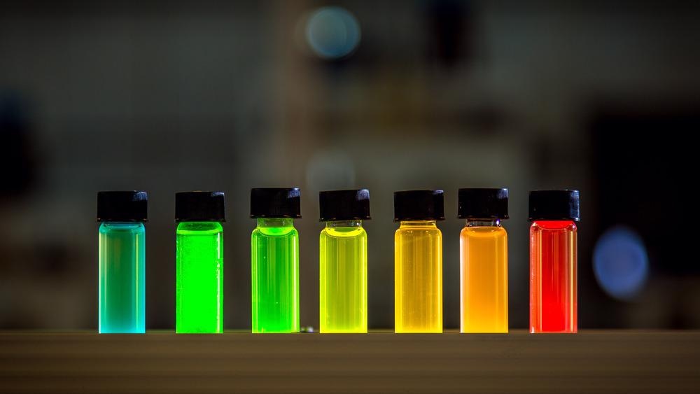
The invention of a rapid, simple, and efficient technique for developing graphene quantum dots utilizing electron beam illumination is the subject of recent research published in the journal Nanotheranostics.

Study: Graphene Quantum Dots prepared by Electron Beam Irradiation for Safe Fluorescence Imaging of Tumor. Image Credit: Tayfun Ruzgar/Shutterstock.com
Due to their unique features in medical applications such as bioimaging, scanning, and drug delivery, graphene quantum dots (GQD) have received a lot of interest in the last decade. However, scale preparation of red luminescing GQD remains a significant challenge.
Graphene Quantum Dots for Medical Applications
Cancer is a dangerous illness that poses a significant threat to human health and wellbeing. Early detection of cancer is crucial to reduce cancer fatalities, increase survival rates, and extend a patient's life.
As a type of quasi-zero-dimensional nanocomposite, quantum dots break past the dimensional barrier, exhibiting a high yield of luminous photons, robust optical characteristics, and light absorption protection compared to standard diagnostic techniques.
Graphene quantum dots (GQD), a unique fluorescent nanomaterial, have a wide range of applications in cell imaging, bioelectronics, and drug delivery devices due to their excellent properties such as high illuminance, durability, oxidative barrier properties, and ease of chemical treatment.

Schematic diagram of irradiation synthesis of graphene quantum dots for tumor fluorescence imaging. © Cao, H. et al. (2022).
Limitations of Existing GQD Production Methods
The aqueous cutting technique, the oxidation cutting carbon fiber method, and the electrolytic peeling method are the most common GQD preparation techniques.
The aqueous cutting technique is comparable to the oxidation cutting carbon fiber process, and although it is a traditional method for making GQD, it is time-consuming and requires the use of a range of strong acids throughout the manufacturing phase. The electrolytic stripping technique has a lengthy pre-processing time, a sluggish post-processing product process, and poor manufacturing efficiency.
As a result, developing a simple, speedy, large-scale, and economic GQD preparation process remains a major issue for researchers.
Radiation Technology for Fabrication of GQD
The benefits of radiation manufacturing processes, such as their quick, efficient, and pure industrial possibilities, have attracted a lot of interest. Because the electron is a highly powerful reducing agent, no additional molecular reductant is required throughout the catalytic process. Furthermore, by adjusting the radiation dosage, the shape and size of the GQD may be altered, thereby providing a way to manufacture small-scale GQD.
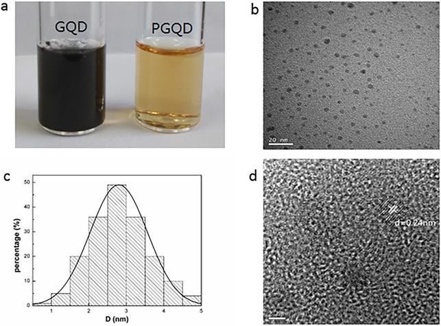
Therefore, the irradiation synthesis technology was effectively used in this work to create red fluorescence GQDs with a size of 2.75 nm. Further functionalization of GQD was accomplished using polyethylene glycol (PEG), which resulted in excellent stability and cytocompatibility.

(a) 2 g/L GQD and PGQD solution. (b) TEM image. (c) Size distribution. (d) HR-TEM image of PGQD. © Cao, H. et al. (2022).
RBC test and Fluorescence Imaging of Tumor
For the red blood cell test, fresh blood was drawn from mice and spun at 4000 rpm for 5 minutes. The blood was then spun again for another 5 minutes.
The plasma was eliminated from the body and the isolated red cells were centrifuged three times in 10 liters of PBS to remove any remaining debris. Following this, the impact of nanomaterials on red blood cells (RBC) was studied.
The optical microscope and scanning electron microscopy photos demonstrate that the structure of RBCs does not change after treatment with 100 g/mL of GQD/PGQD for 4 hours when compared with the control, indicating that the cytotoxicity of GQD and PGQD to RBCs was not highly significant.
At 610 nm, PGQD might produce a bright red luminescence. Mice with 4T1 tumors were given PGQD dispersion solution orally and scanned using a small mammal fluorescence imaging technology.
Heavy red fluorescent signals were found in the tumor 5 minutes after administration, showing that PGQD would preferentially and quickly concentrate in the tumor region. The fluorescent signals in the tumor decreased with time, demonstrating that tiny PGQD may be removed over time.
These findings demonstrate that PGQD produced by electron beam irradiation may be used as a tumor-targeting diagnostic reagent.

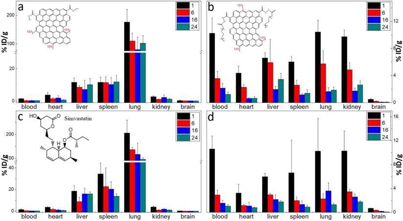
The biodistribution of GQD (a) and PGQD (b) the effect of simvastatins on the biodistribution of GQD (c) and PGQD (d) in mice at 1, 6, 16 and 24 h post injection of 131I-labeled materials. © Cao, H. et al. (2022).
Research Conclusions
Using electron beam irradiation, graphene quantum dots that were consistent in size and good luminous properties were effectively synthesized.
GQDs were further synthesized and characterized with PEG and showed high stability and cytocompatibility.
PEGylated GQD was shown to preferentially concentrate in tumors following intramuscular infusion, suggesting that it might be used as a sensitive tumor optical sensing agent. This paper described a novel technique for making red-luminescing GQD for biological use.
Continue reading: Recent Advances and Perspectives of DNA-Nanosensors for Environmental Monitoring.
Reference
Cao, H. et al. (2022). Graphene Quantum Dots Prepared by Electron Beam Irradiation for Safe Fluorescence Imaging of Tumor. Nanotheranostics, 6(2), 205-214. Available at: https://www.ntno.org/v06p0205.htm
Disclaimer: The views expressed here are those of the author expressed in their private capacity and do not necessarily represent the views of AZoM.com Limited T/A AZoNetwork the owner and operator of this website. This disclaimer forms part of the Terms and conditions of use of this website.