Drugmakers have been using genetically modified cells as small drug factories for over 40 years. These cells can be instructed to produce chemicals that can be utilized to treat cancer and autoimmune diseases like arthritis.
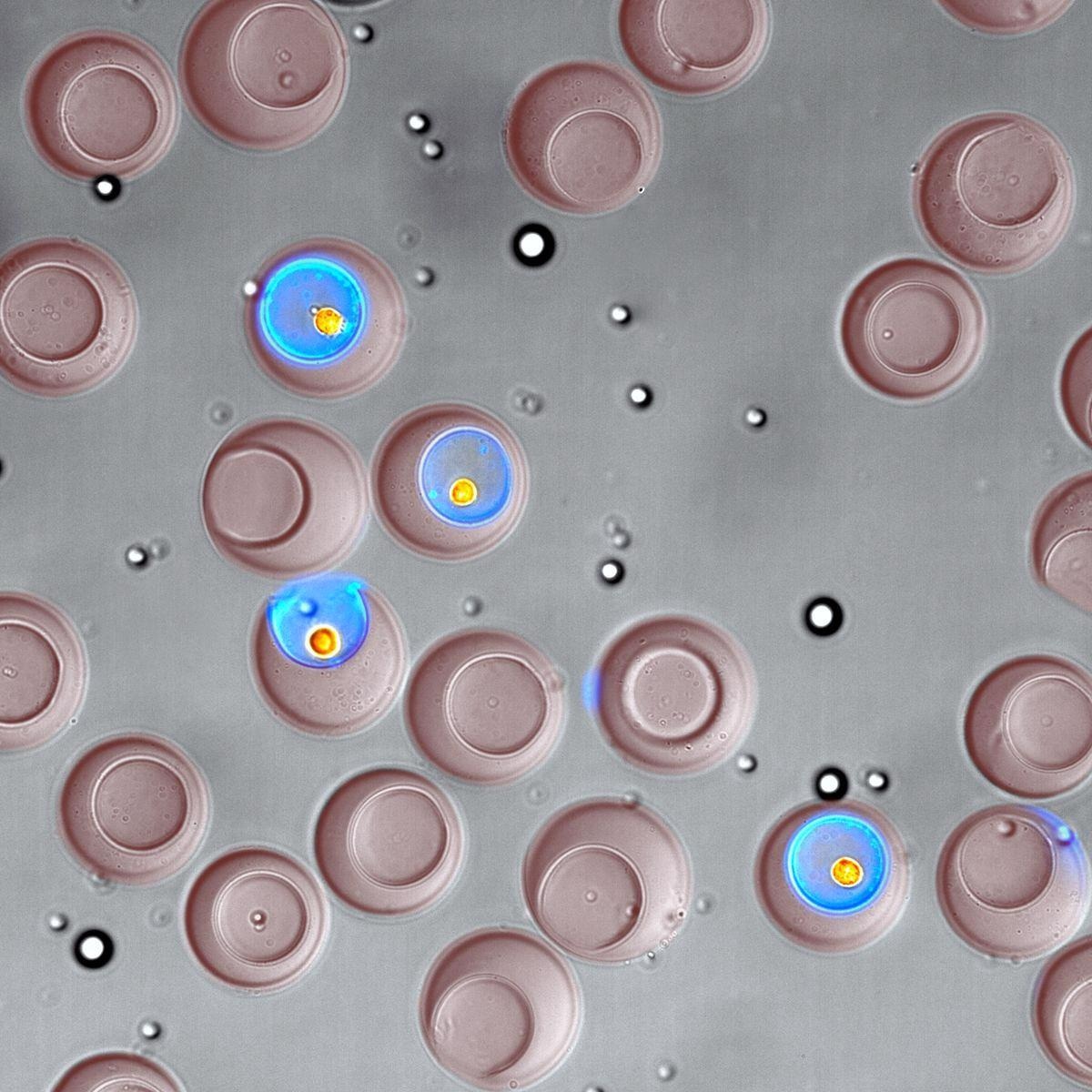
 Using microscopic, bowl-shaped containers called nanovials (the larger, reddish-brown objects) enabled researchers to select cells based on what type they are and which compounds they secrete (shown here in blue). Image Credit: Joseph de Rutte/ The University of California, Los Angeles.
A novel approach for efficiently sorting single living cells in a typical laboratory setting might help with efforts to design and produce new biologic treatments. A UCLA-led research team recently showed the capacity to choose cells depending on their kind and which substances — and how much of those compounds — they produce using microscopic bowl-shaped hydrogel containers called “nanovials.” The research was reported in the journal ACS Nano.
Basic biological research might potentially benefit from the technology.
With this technology, the scientific community can uncover new insights into key biological processes that represent a huge fraction of our protein-coding genes. I think of the single cell as the quantum limit of biology. The nanovial is the evolution of the Petri dish to that fundamental limit of one cell.
Dino Di Carlo, Study Corresponding Author and Armond and Elena Hairapetian Professor, Engineering and Medicine, Samueli School of Engineering, University of California
Nanovials, according to Di Carlo, enable scientists to overcome the limits of traditional devices for detecting cell secretions. Di Carlo is also a member of the California NanoSystems Institute at UCLA and the UCLA Jonsson Comprehensive Cancer Center.
The most frequent method uses a microwell plate, which consists of a grid of small plastic containers. However, this method lacks the nanovial’s capacity to sort single cells, and current technology often takes weeks to grow enough cells to detect secretions. The second option is a multimillion-dollar instrument that monitors the secretions of around 10,000 cells per experiment and can sort living cells, but it is only found in a few dozen labs across the world.
Nanovials, in comparison to that device, can do much larger screenings — millions of cells — at a fraction of the cost — less than one cent per cell, vs $1 or more per cell with the present standard.
Nanovials are so small that to fill a teaspoon, 20 million of them would be needed. They may be tailored to catch certain cell types and enhanced with molecules that adhere to a cell’s secretions and glow in different colors when exposed to light. As nanovials are constructed of a hydrogel, which is a polymer that can hold about 20 times its weight in water, they create a wet environment that is comparable to that seen in cells’ natural environments.
Researchers looked at cells that had been genetically modified to release a specific antibody drug. Scientists picked cells that produced the most of that antibody using nanovials and a common analytical instrument called a flow cytometer, and then expanded those cells into colonies that generated nearly 25% more of the medication than colonies that had not been carefully selected.
The capacity to make antibody drugs with that level of efficiency might cut medication production costs by a similar amount, according to Di Carlo.
The researchers also showed that they could detect rare antibody-secreting cells that bonded exclusively to a target molecule, as well as the secreted antibody’s DNA sequence information.
Nanovials are now being utilized by researchers to examine immune cells called T cells, which are used in cell treatments, as well as to investigate previously unknown biological processes. Nanovial technology is also the foundation for Partillion Bioscience, a venture established at CNSI’s Magnify incubator on the UCLA campus.
We are excited to see the impact nanovials will have for biological research now that they are available for anyone to use.
Joseph de Rutte, Study Co-First Author and Doctorate, University of California
Joseph de Rutte is also the co-founder and president of Partillion.
The paper’s other co-first author is Robert Dimatteo, who received his doctorate from UCLA in 2021.
Maani Archang, Sohyung Lee and Kyung Ha, who recently received doctorates from UCLA; present doctoral students Mark van Zee, Doyeon Koo, Michael Mellody and Shreya Udani; assistant project scientist Allison Sharrow; James Eichenbaum, who recently obtained a bachelor’s degree; and professors Andrea Bertozzi and Robert Damoiseaux are among the other UCLA co-authors. Johns Hopkins University and the University of Houston are among the other authors.
The National Institutes of Health, the Maryland Stem Cell Research Fund, and the Simons Foundation all contributed to the research.
Journal Reference:
de Rutte, J., et al. (2022) Suspendable Hydrogel Nanovials for Massively Parallel Single-Cell Functional Analysis and Sorting. ACS Nano. doi.org/10.1021/acsnano.1c11420.