A new paper from the ACS Nano demonstrated the feasibility of using a modular biotin-streptavidin assembly of nanocomposites for intracellular cytosolic protein delivery.

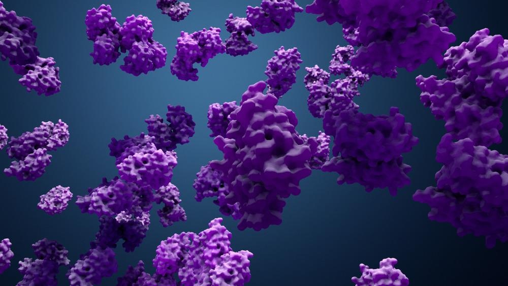
Study: Cytosolic Protein Delivery Using Modular Biotin–Streptavidin Assembly of Nanocomposites. Image Credit: Design_Cells/Shutterstock.com
Importance and Challenges of Intracellular Protein Delivery
Intracellular protein delivery represents a revolutionary technology for fundamental research and biomedical applications. Protein plays a crucial role in homeostasis and cell signal transduction. Moreover, several inherited diseases are caused by abnormal protein function.
Although over 130 Food & Drug Administration (FDA)-approved protein therapeutics are available in the market, the effectiveness of these therapeutics has been limited to extracellular targets owing to their cell membrane impermeability.
To achieve cytosolic protein delivery, strategies need access to intracellular targets, such as the nucleus and subcellular organelles. A significant share of existing intracellular protein delivery approaches depends on an endosomal uptake of nanovectors or modified proteins.
Endosomal entrapment of proteins can severely limit the effectiveness of intracellular delivery strategies by preventing access to the cytosol. Additionally, protein cargo entrapped in endosomes is often degraded through the lyso/endosomal pathway or exocytosed, making endocytosis a challenging route for intracellular protein delivery.
Potential Ways to Achieve Intracellular Protein Delivery
Typically, less than ten percent of endocytosed protein escapes into the cytosol through existing delivery approaches, which can be increased by engineering delivery vehicles that can trigger endosomal rupture and facilitate the escape of proteins into the cytosol. The fusion of protein delivery vectors with the cell membrane can provide a direct mechanism of intracellular delivery, bypassing the issues related to endosomal entrapment.
Versatile delivery platforms such as polymer scaffolds act as a suitable intracellular delivery vehicle for proteins as they provide design flexibility through chemical diversity. For instance, cationic fluoropolymers or dendrimers were utilized to achieve cytosolic protein delivery for extensive applications such as therapeutic gene editing and cancer immunotherapy.
Previous studies have demonstrated direct cytosolic protein delivery using co-engineered proteins modified with terminal oligo (glutamate) “E-tags” and guanidinium-functionalized gold nanoparticles. Recently, the strategy was adapted for guanidinium-functionalized poly(oxanorbornene) imide (PONI-Guan) polymers to achieve highly effective cytosolic delivery. However, both strategies require plasmid engineering to obtain E-tagged proteins different from native proteins, which makes these strategies cumbersome.
New Strategy Proposed for Cytosolic Protein Delivery
Tetravalent biotin-streptavidin (STV) binding offers a modular high-affinity strategy for bioconjugation of protein. Different macromolecular and molecular systems can be biotinylated, making the STV an extremely versatile platform for protein bioconjugation.
In this study, researchers used modular streptavidin-biotin assemblies of nanocomposites to achieve cytosolic delivery of proteins. Initially, biotinylated proteins, including biotinylated green fluorescent protein (b-GFP), and biotinylated oligo(glutamate) (b-E20) were conjugated to STV.
Conjugates were then self-assembled using the PONI-Guan homopolymer to obtain discrete supramolecular protein-polymer nanocomposites. GFP was utilized as a model protein owing to its ability to passively diffuse into the nucleus and robust fluorescence. Thus, it can provide a clear indication of cytosolic access.
Synthesis, Characterization, and Evaluation of Biotinylated Proteins and Nanocomposites
b-GFP was synthesized by amine-reactive crosslinking of wild-type GFP through the N-hydroxysuccinimide (NHS) ester coupling. b-E20 and additional biotinylated proteins were also synthesized similarly as b-GFP.
PONI-Guan polymer/STV/b-E20/b-GFP nanocomposites were synthesized in polypropylene microcentrifuge tubes. Initially, STV was added to a mixture of b-GFP and b-E20 by pipet and the resultant mixture was incubated for ten minutes at an ambient temperature. Subsequently, PONI-Guan homopolymer was added to the nanocomposite mixture, and the mixture was again incubated for ten minutes at ambient temperature to obtain the final PONI-Guan polymer/STV/b-E20/b-GFP nanocomposites.
Transmission electron microscopy (TEM) was used to characterize the synthesized samples, while cytosolic delivery and effective nuclear access of b-GFP were quantified using imaging flow cytometry. Electrospray ionization mass spectrometry and confocal microscopy imaging were used to verify biotinylation and assess cytosolic delivery through fluorescence, respectively.
Additionally, colorimetric 4′-hydroxyazobenzene-2-carboxylic acid (HABA) displacement assay was utilized to confirm the binding between b-E20, b-GFP, and STV. Finally, the retention of protein function after cytosolic delivery was evaluated by delivering biotinylated granzyme A (b-GrA), a chemotherapeutic enzyme.
Research Findings
PONI-Guan/STV/b-E20/b-GrA nanocomposites were synthesized successfully with an average size between 200 nanometers and 350 nanometers. The HABA displacement assay confirmed the binding between the proteins and STV. All nanocomposites possessed a spherical morphology and a similar size.
Effective cytosolic delivery of b-GFP proteins in both human cervix adenocarcinoma and hamster ovary epithelial cells was achieved through the nanocomposites. In addition to b-GFP, effective cytosolic delivery was also achieved for the additional proteins, demonstrating the biotin-streptavidin-based tethering strategy for intracellular protein delivery.
In confocal microscopy, diffused green fluorescence was observed throughout the cytosol, indicting cytosolic delivery. The cytosolic delivery of protein occurred within three to five hours of initial incubation of the nanocomposites. No significant toxicity and changes in cell morphology were observed after incubation.
The retention of protein function after the cytosolic delivery was confirmed through the efficient killing of cells after the delivery of the GrA. The cytosolic delivery of GFP displayed a greater efficiency when the nanocomposites were used as delivery vectors compared to the commercially available protein delivery reagents.
To summarize, the findings of this study demonstrated that PONI-Guan/STV/b-E20/b-GrA nanocomposites represent a versatile and modular approach to intracellular protein delivery through the noncovalent tethering of several components into a single delivery vehicle.
Reference
Goswami, R., Gopalakrishnan, S., Nagaraj, H. et al. (2022) Cytosolic Protein Delivery Using Modular Biotin−Streptavidin Assembly of Nanocomposites. ACS Nano. https://pubs.acs.org/doi/10.1021/acsnano.1c06768
Disclaimer: The views expressed here are those of the author expressed in their private capacity and do not necessarily represent the views of AZoM.com Limited T/A AZoNetwork the owner and operator of this website. This disclaimer forms part of the Terms and conditions of use of this website.