A group of researchers recently published a paper in the journal Scientific Reports that demonstrated the effectiveness of gold nanoparticles synthesized using hydrogels as reducing and capping agents for wound healing and antimicrobial applications.

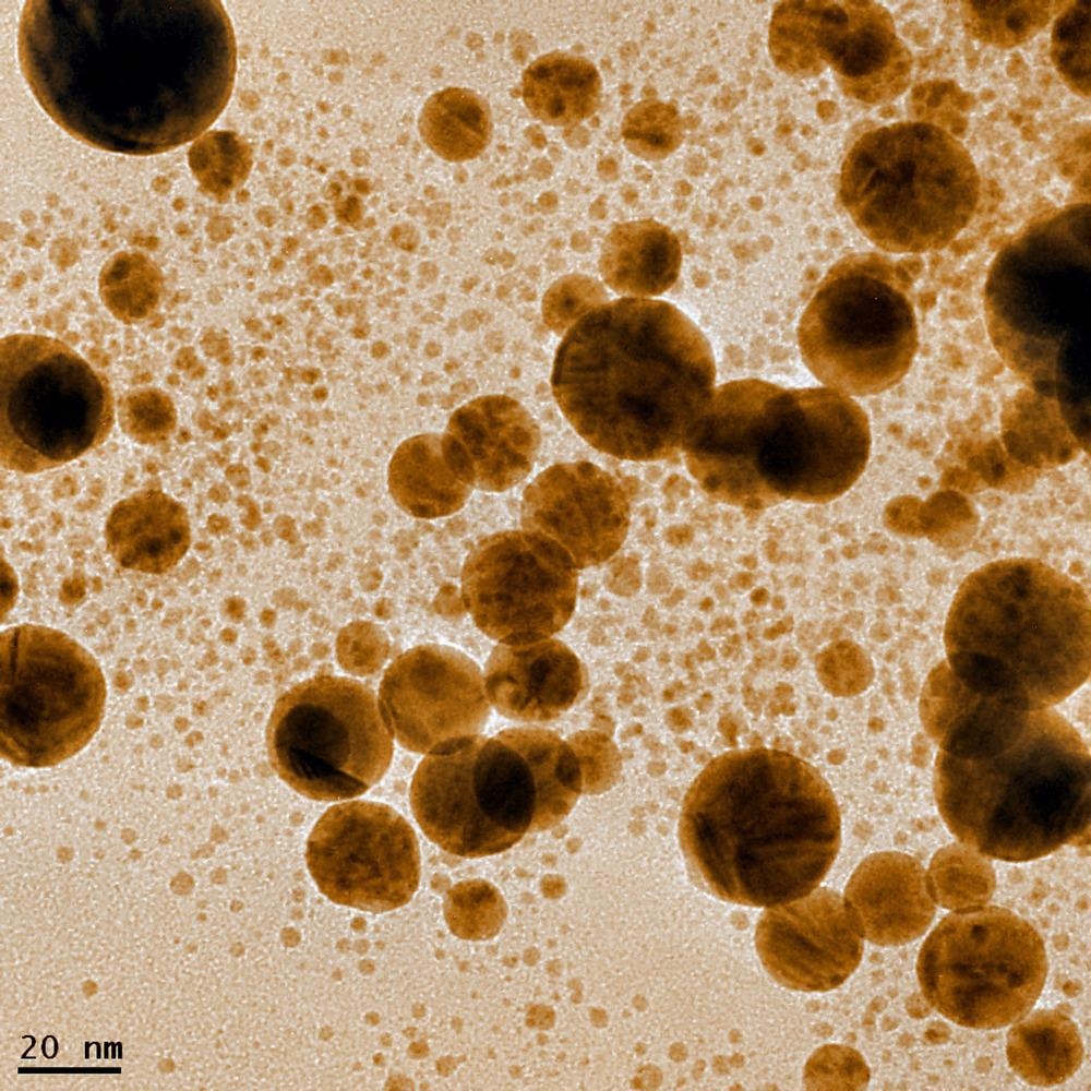
Study: Hydrogel assisted synthesis of gold nanoparticles with enhanced microbicidal and in vivo wound healing potential. Image Credit: Georgy Shafeev/Shutterstock.com
Importance of Green Synthesis of Gold Nanoparticles
Nanoparticles possess superior microbicidal properties as they can penetrate the cell walls and cell membranes of pathogens more easily compared to the common anti-bacterial and anti-fungal drugs. For instance, the antimicrobial characteristics of polymer-based wound dressing can be enhanced significantly by incorporating selenium and gold-based nanoparticles into the nanofibers.
Gold nanoparticles are considered more suitable for biomedical applications compared to other metallic nanoparticles owing to their non-toxic nature. Gold nanoparticles can be prepared by the reduction of gold (III) salts using hydrogen peroxide and gallic acid as reducing agents.
However, certain reducing agents, such as hexadecyltrimethylammonium bromide and sodium borohydride, are considered perilous and toxic to the environment, which necessitated the development of eco-friendly, biocompatible, and clean methodologies for nanoparticle synthesis.

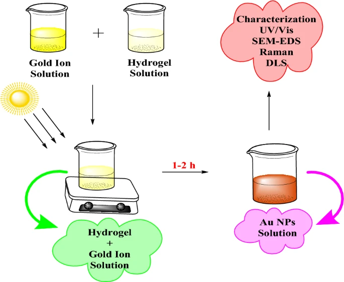
Schematic representation of the sunlight-assisted synthesis of Au NPs. Image Credit: Syed, A et al., Scientific Reports
The green nanoparticle synthesis methods that utilize plants, algae, fungi, and bacteria during the synthesis are considered economical and eco-friendly. These synthesis methods often use hydrogels and other natural products present in living organisms as capping and reducing agents.
Hydrogels have attracted significant attention as reducing agents for nanoparticle synthesis owing to their biocompatibility, biodegradability, and availability. Additionally, hydrogels can absorb a substantial amount of aqueous solutions owing to the presence of functional groups such as carboxyl and hydroxyl groups.
Although hydrogels were used for the synthesis of silver nanoparticles, there is no instance of using them in the preparation of gold nanoparticles.
Synthesis, Characterization, and Evaluation of Gold Nanoparticles
In this study, researchers proposed a sunlight-assisted hydrogel-based green synthesis method for synthesizing gold nanoparticles. The hydrogel extracted from Cydonia oblonga (C. oblonga) seeds was used as the capping and reducing agent for gold nanoparticle synthesis for the first time.
Initially, C. oblonga seeds were soaked in water for 20 minutes at room temperature, and then a cotton muslin cloth was used to separate the hydrogel extracted from those seeds. Subsequently, the hydrogel was dehydrated for four to five hours at 60-65 degrees Celsius in a hot air oven and ground into a fine powder. Hydrogen tetrachloroaurate (III) was used as a gold precursor.

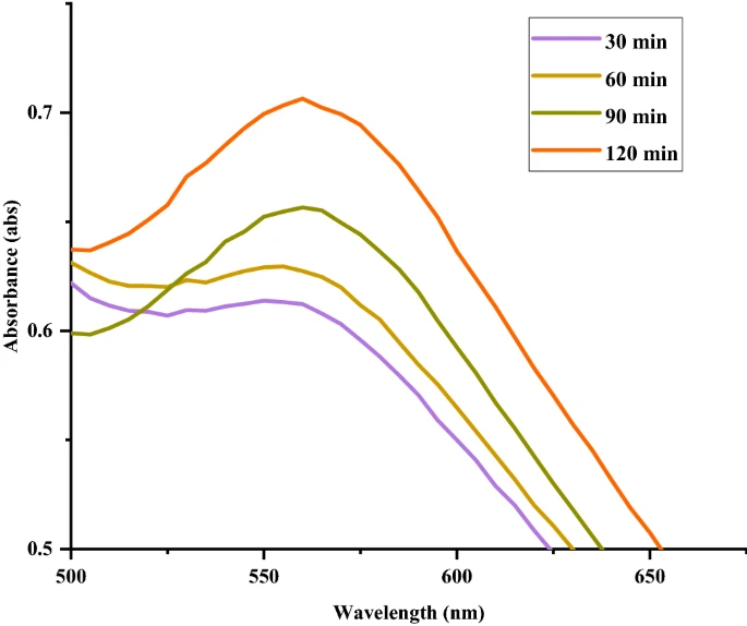
UV–Vis spectra show the changes in absorption of Au NPs at different times during synthesis. Image Credit: Syed, A et al., Scientific Reports
The synthesized hydrogel powder was suspended in deionized water to obtain the hydrogel suspension. The gold precursor solution was also prepared in a similar manner by dissolving the hydrogen tetrachloroaurate (III) in deionized water. Equal amounts of hydrogel suspension and gold precursor solution were mixed and stirred at room temperature.
The formation of gold nanoparticles in the reaction upon exposure to sunlight was monitored by color variation. Additionally, the ultraviolet-visible (UV-Vis) spectra were also obtained at different stages of synthesis using a UV–Vis spectrophotometer to monitor the localized surface plasmon resonance (LSPR) absorption bands to further confirm the formation of gold nanoparticles.
The synthesized gold nanoparticles were characterized using dynamic light scattering (DLS), transmission electron microscopy (TEM), X-ray diffraction (XRD), Raman spectroscopy, UV-Vis spectroscopy, and scanning electron microscopy (SEM). Researchers also evaluated the anti-bacterial and anti-fungal activities and in vivo wound healing activity of gold nanoparticles.
The fungicidal and bactericidal activities of the synthesized gold nanoparticles were analyzed against fungal strains Penicillium notatum and Aspergillus niger and bacterial strains Staphylococcus aureus, Pseudomonas aeruginosa, Escherichia coli, Bacillus subtilis, and Bacillus simplex, respectively. The quantitative analysis of biomarkers related to wound healing was performed using the CYBER green real-time polymerase chain reaction (PCR) kit.
Research Findings
Gold nanoparticles were synthesized successfully using the light-assisted green synthesis method. Upon exposure to sunlight, the color of the reaction changed from yellow to reddish-brown due to the reduction of gold ions into nanoparticles, which confirmed the formation of gold nanoparticles. Moreover, the UV-Vis spectra of synthesized gold nanoparticles displayed an SPR absorption band at 560 nanometers, which corresponds to the LSPR of typical gold nanoparticles.
The XRD pattern of gold nanoparticles confirmed the cubic structure of gold nanoparticles. The average edge length and size of rectangular and cubic-shaped gold nanoparticles were 74± 4.57 nanometers and 20-30 nanometers. The gold nanoparticles were stabilized by the functional groups present in the hydrogel. Raman spectroscopy confirmed the successful gold nanoparticle formation with homogenous size and capping of hydrogel functional groups with gold nanoparticles. The particle size distribution of most of the gold nanoparticles was 25 nanometers.

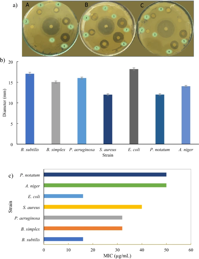
The anti-microbial potential of Au NPs: (a) Zones of inhibition of model bacterial strains (A) B. simplex, (B) E. coli, (C) S. aureus; 1: Ampicillin; 2–7: 10–50 µL/mL Au NPs. (b) Graph showing maximum diameter for zones of inhibition of various microbial strains. The error bars show the standard deviation. (c) Graph showing MIC values of various microbial strains. Image Credit: Syed, A et al., Scientific Reports
The synthesized gold nanoparticles successfully prevented bacterial and fungal growth within 24 hours. The gold nanoparticles inhibited bacterial growth with inhibition zones between 12 and 18 millimeters and fungal growth with inhibition zones between 12 and 14 millimeters in diameter.
In vitro studies showed that the minimum inhibitory concentration (MIC) values of gold nanoparticles against the bacterial strains were between 16 and 40 micrograms per milliliter, and 50 micrograms per milliliter against fungal strains. The MIC value considered the gold nanoparticle concentration that reduced 95 percent growth of fungal and bacterial strains.
In vivo studies in murine models demonstrated that 90 percent of the wound treated with gold nanoparticles healed within five days. Quantitative PCR analysis showed that the expression levels of CD-34 and NANOG proteins increased significantly in gold nanoparticle-treated wound tissues, while the expression level of MMP-2 decreased.
To summarize, the findings of this study demonstrated that gold nanoparticles synthesized using hydrogels as reducing agents possess significant in vivo wound healing, antimicrobial, and antifungal properties. Thus, these NPs can be used effectively in biomedical applications such as cancer treatment and controlled drug delivery.
Disclaimer: The views expressed here are those of the author expressed in their private capacity and do not necessarily represent the views of AZoM.com Limited T/A AZoNetwork the owner and operator of this website. This disclaimer forms part of the Terms and conditions of use of this website.
Source:
Syed, A., Sajjad, N., Batool, Z. et al. Hydrogel assisted synthesis of gold nanoparticles with enhanced microbicidal and in vivo wound healing potential. Scientific Reports 2022. https://www.nature.com/articles/s41598-022-10495-3