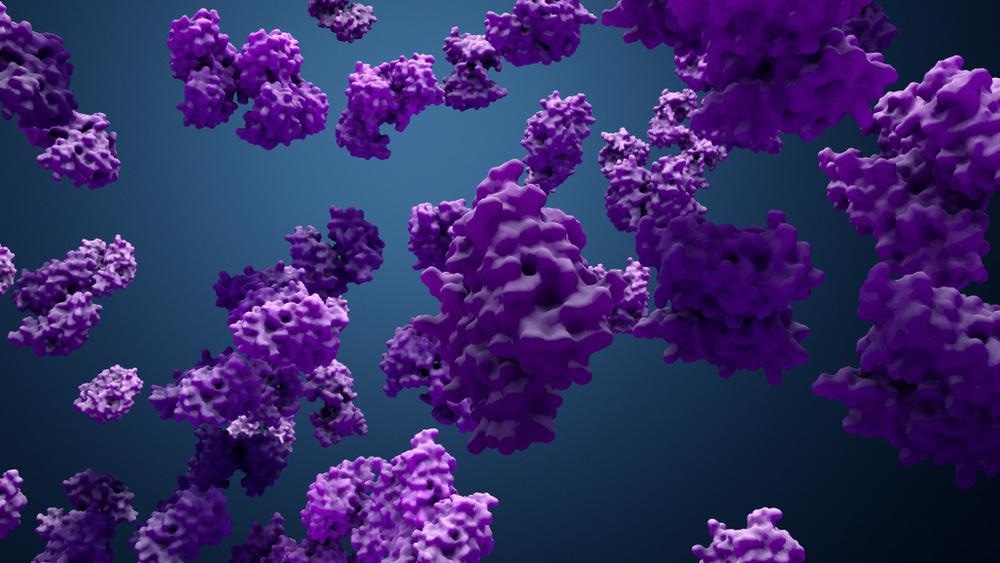
Cryogenic transmission electron microscopy (cryo-EM) is one of the recent methods for obtaining high-resolution images of proteins. However, this technique comes with certain limitations. Researchers developed giant carbon nano-test tubes (GCNTTs) as radiation-insulating material to address this, which helps procure high-resolution protein images without affecting the sample. This study is available in ACS Applied Materials and Interfaces.

Study: Giant Carbon Nano-Test Tubes as Versatile Imaging Vessels for High-Resolution and In Situ Observation of Proteins. Image Credit: Design_Cells/Shutterstock.com
Protein Imaging Techniques
Cryo-EM, coupled with image analysis techniques, has significantly contributed to protein structure databases. Although many advancements are found related to the resolution and technical aspects of protein imaging, not many improvements are reported in the sample preparation area.
Both X-ray crystallography and cryo-EM require protein samples to be in a frozen state for analysis. This kind of sampling system limits the ability to gather dynamic and functional information; thereby, scientists fail to understand proteins in physiological conditions.
Although standard electron microscopy can procure structural details of inorganic compounds, it exhibits low resolution for biological samples due to irreversible damage of the specimen via irradiation. To overcome this limitation, scientists have developed new materials, for example, single-walled carbon nanotubes (SWCNTs), that can protect biomolecules from degradation by the electron beam.
Several studies have shown that SWCNT has unique thermal and electrical properties, which are highly advantageous for EM-based imaging methods. However, this method is restricted to small molecules due to its tiny pore size. Thereby, the synthesis of carbon nanotubes, with larger pore size, with radiation insulation properties could be applied for EM-based techniques to capture high-resolution images of larger biomolecules with complex structures.
Giant Carbon Nano-test Tubes for High Resolution In situ Observation of Proteins - A New Study
Scientists stated that the development of new carbon nanotubes with larger pore sizes with irradiation insulation properties would be highly beneficial for protein imaging studies, which had been difficult using conventional techniques, e.g., X-ray crystallography and static cryo-EM techniques. The new technique presents a unique opportunity to conduct real-time, unstained protein imaging under dynamic and static environments.
In this study, researchers have developed GCNTTs by depositing carbon on anodized aluminum oxide (CAAO) templates, with tunable nanopore sizes varying between 20 and 200 nm. The depths of the pores could be customized between 500 nm and tens of microns to fit the biomolecules of interest. As hemoglobin (Hb) and fructose dehydrogenase (FDH) are the target molecules of this study, the pore size of GCNTTs was maintained at around 30 and 60 nm, respectively.
Scientists observed that GCNTTs synthesized by the CAAO method possessed similar longitudinal thermal conductivity as SWCNTs developed via conventional methods.
Characterization of newly developed GCNTTs using transmission electron microscopy (TEM) revealed a wall thickness of 1−3 nm and inner diameters of approximately 30 and 60 nm. In this study, researchers mixed GCNTTs into a Hb solution and the proteins were allowed to bind passively with GCNTTs. Subsequently, they characterized the GCNTT-Hbs using TEM and observed that the proteins were easily located because Hbs were spatially constricted within the newly synthesized GCNTT with a larger pore diameter. Scientists found that the proteins were lined, regularly spaced within the inner wall of GCNTT, and were approximately 50 nm in length.
In this study, researchers could image Hb embedded within GCNTT via TEM, without requiring additional sample preparation steps which are required for X-ray crystallography or cryo-EM. Additionally, scientists lyophilized GCNTT-Hb samples directly onto the TEM grid to analyze the possibility to image Hb molecules in a dispersive state within GCNTT. Standard TEM analysis revealed that ∼10 nm spherical, evenly spaced particles were dispersed within GCNTT, whose diameter (7 nm) was similar to native Hb. Hence, researchers believe that Hb molecules were monodispersed.
Scientists imaged the GCNTT-Hbs with transmission electron microscopy (TEM) and scanning electron-assisted dielectric impedance microscopy (SE-ADM) to determine that the high-power electron beam does not irradiate and damage the protein samples embedded within the GCNTT.
In this study, the dispersed GCNTT-Hb sample was exposed to 50,000× magnification and 3 kV irradiation. Interestingly, no change in Hb biomolecule was observed, indicating that GCNTT had successfully shielded Hb proteins from the electron beam. Additionally, scientists used scanning TEM (STEM) analysis to study the spatial distribution of each Fe atom within the Hb to demonstrate that the protein sample was not degraded.
Researchers also reported that GCNTT could be applied for fluorescence bleaching studies, which utilize light-sensitive fluorophores or biological molecules sensitive to degradation. Importantly, the authors have successfully studied FDH using the GCNTT platform using TEM analysis.
Future Perspective
This study revealed that GCNTT is a powerful vessel for imaging biomolecules and protecting them from damage from electron beams. In the future, the authors aim to study if the GCNTT platform could be effective for studying other proteins as well. If so, it would present an excellent opportunity to elucidate the surface functionalization and binding kinetics of various proteins.
Reference
Chuong, T.T. et al. (2022) Giant Carbon Nano-Test Tubes as Versatile Imaging Vessels for High-Resolution and In Situ Observation of Proteins. ACS Applied Materials and Interfaces. https://pubs.acs.org/doi/10.1021/acsami.2c06318
Disclaimer: The views expressed here are those of the author expressed in their private capacity and do not necessarily represent the views of AZoM.com Limited T/A AZoNetwork the owner and operator of this website. This disclaimer forms part of the Terms and conditions of use of this website.