Pulsed laser ablation in liquid media (PLAL) is a green technique that allows the formation of nanomaterials with high purity. It is a physical alternative to traditional chemical methods. In an article recently published in the journal Scientific Reports, researchers discussed the preparation of gold nanoparticles (AuNPs) in Gum Arabic (GA) solution, where laser ablation was used as computed tomography (CT) contrast agent.

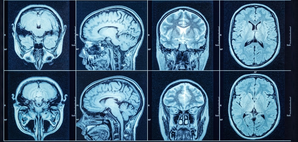
Study: Green synthesis of gold nanoparticles in Gum Arabic using pulsed laser ablation for CT imaging. Image Credit: Triff/Shutterstock.com
The team evaluated the optical properties of synthesized NPs by employing the absorption spectroscopy technique. The information on the shape and size of the prepared nanoparticles was obtained from transmission electron microscope (TEM) images and ImageJ software. TEM images revealed that GA-AuNPs had a spherical shape with good stability and their size was smaller than AuNPs without GA.
The GA-AuNPs absorption spectrum showed a blue shift towards a lower wavelength with a higher plasmon peak height at 514 nanometers. With the help of the present work, the researchers demonstrated the positive influence of GA on AuNPs.
Nanomaterials in CT Imaging
Nanomaterials have unique physical properties and are a robust tool for biomedical applications. Metal NPs are utilized in the fields of medicine, biosensing, food, cosmetics, and biomedical sciences.
AuNPs are non-toxic in nature, easily available, shape and size-controlled, and have the capacity for surface modification with high particle reactivity. Owing to AuNP’s localized surface plasmon resonance (LSPR), AuNPs show purple or red color at a wavelength of 530 nanometers. Additionally, the properties of the AuNPs are modified by controlling the shape and size or through surface modification via a synthetic process.
The synthesized NPs can be surface functionalized by introducing ligands. This functionalization enhances their hydrophilicity and stability, thus preventing the collision and subsequent agglomeration of NPs.
To functionalize the nanoparticles, the ligands are added during their synthesis. Furthermore, the media in which NPs are synthesized have a critical role in functionalizing them. The ionic interactions between media and functionalizing agents may lead to NP instability resulting in their aggregation.
PLAL involves solid target ablation by employing strong laser rays resulting in the production of nanomaterials. PLAL is a cost-effective and non-toxic method that allows the production of nanomaterials with high purity. This method can be employed to obtain AuNPs without reducing agents.
Computed tomography (CT) is a diagnostic approach that uses X-rays in high-resolution 3D anatomic imaging. However, X-ray attenuation has a tiny difference for soft tissues, which makes it difficult to distinguish between surrounding tissues. Although iodine contrast can overcome these limitations, nanomaterials are better contrast agents.
GA-AuNPs in CT Imaging
In the present study, the researchers aimed to use GA-AuNPs in CT imaging as a contrast agent and explore their properties. They employed the PLAL method to synthesize GA-AuNPs using lasers of different energies. They investigated the variation in particle size by varying the GA concentrations and number of laser pulses.
AuNPs were synthesized in deionized water in the presence and absence of GA and by employing laser ablation. The researchers observed a color change after seconds of ablation. The AuNP sample appeared light red, and GA-AuNPs were dark purple. This color change was due to LSPR.
Atomic absorption spectroscopy (AAS) revealed that the concentration of GA-AuNPs was higher than AuNPs due to the GA coating that avoided aggregation of AuNPs. On the other hand, naked AuNPs aggregated to form particles of larger size and consequently reduced the number of particles.
The absorbance at high laser energy showed an increase in the height of the plasmon peak, indicating no significant change in AuNP’s concentration. With an increase in energy, the peak showed a blue shift. Additionally, at high energies, the NPs showed an initial greater distribution and average size. However, with continuous laser irradiation, the size and distribution showed a reducing trend due to fragmentation.
Conclusion
In summary, the researchers illustrated the synthesis of AuNPs using the PLAL technique. They observed that the presence of GA significantly impacted the size and production of NPs. Compared to AuNPs, the GA-AuNPs had a smaller size with no aggregation because GA acted as a stabilizing agent that controlled the AuNP’s production and coating.
Twenty milligrams of GA achieved a blue shift with an absorption peak at 514 nanometers, providing the NPs a higher stability and size reduction. Laser ablation parameters hugely impacted the properties of GA-AuNPs. Moreover, the number of laser pulses influenced the qualities of GA-AuNPs.
Energy greater than 200 millijoules provided high stability and smaller sizes to NPs. The concentration of GA-AuNPs affected the quality of the CT image. Further, increasing GA-AuNPs concentration to 54 parts per million improved the image contrast by enhancing the CT number.
Based on these findings, the researchers concluded that GA could serve as a promising contrast agent for biomedical applications such as medical imaging and magnetic resonance imaging (MRI), and as a targeting agent for cancer therapy.
Reference
Mzwd, E., Ahmed, N.M., Suradi, N., Alsaee, SK., Altowyan, AS., Almessiere, MA., Omar, AF et al. (2022) Green synthesis of gold nanoparticles in Gum Arabic using pulsed laser ablation for CT imaging. Scientific Reports. https://doi.org/10.1038/s41598-022-14339-y
Disclaimer: The views expressed here are those of the author expressed in their private capacity and do not necessarily represent the views of AZoM.com Limited T/A AZoNetwork the owner and operator of this website. This disclaimer forms part of the Terms and conditions of use of this website.