In the era of biomedical applications of nanoparticles, it is imperative to determine how they affect biological functions and the fate of nanomedicine. This analysis would positively help optimize nanomedicine, reducing side effects, improving clinical translation, and maximizing its impact.

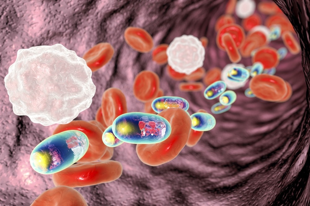
Study: Advanced Light Source Analytical Techniques for Exploring the Biological Behavior and Fate of Nanomedicines. Image Credit: Kateryna Kon/Shutterstock.com
The rapid growth in advanced light source (ALS) analytical technologies has enabled scientists to determine the fate of nanomedicines in vivo. Researchers have recently reviewed ALS analytical technologies, especially spectroscopy and imaging, to highlight their applicability in determining the biological behavior and fate of nanomedicines. This review is available in ACS Central Science.
Factors that Affect the Bioidentity of Nanomedicines
Scientists have discovered many novel nanomedicines that are applied in various areas of biomedicine, including drug delivery, diagnosis, and therapy. Even though nanomedicine applications have immensely improved the efficacy of conventional medicines, researchers face varied difficulties associated with its production, preclinical characterization, and clinical outcomes.
Previous studies have revealed that the physicochemical properties of nanoparticles, i.e., size, shape, and charge, determine the efficacy and fate of nanomedicines. For instance, in vivo formation of protein corona on the surface of nanoparticles alters its biodistribution and characteristic properties, affecting the bioidentity of nanomedicines.
The transformation of nanomedicines within the complex environment of biological systems also induces changes in their surface properties and function. However, the exact physiochemical behavior of nanostructured nanomedicines within the biological environment is not well understood.
Understanding the complex biological functions, the relationship between a nanomaterial and biological component (nano-bio interaction), nanomedicine’s fate, and spatiotemporal interactions among various nanoparticles is important for optimizing nanomedicines.
Information from the in-depth characterization of nanomedicines could help in the rational design of future nanomedicines that would prevent oxidative stress, toxicity, formation of the surface protein corona, and genetic damage.
Analysis of Nanomedicine Behavior in Biological Environments
Several imaging techniques, such as electron microscopy, optical microscopy, and positron emission tomography/single photon emission computed tomography (PET/SPECT), are used to study the behavior of nanomedicines in dynamic biological environments. Although electron microscopy, which includes scanning transmission microscopy (SEM) and transmission electron microscopy (TEM), is used to capture high-resolution images, in situ imaging of internal structure within the cells or tissues is difficult.
Fluorescence microscopy is a type of optical microscopy that enables high-resolution imaging of the dynamic behavior of nanomedicines. However, one of the limitations associated with applying this microscopy is the lack of suitable fluorescence probes. PET/SPECT are used to image humans and small animals; however, this technique requires radiolabelled nanomaterials.
ALS Imaging and Spectroscopic Analysis to Evaluate Nanomedicines in Biological Environments
Recently, ALS imaging and spectroscopic technologies have been used to study nano−bio interactions. ALS imaging technology (e.g., X-ray) enables deep penetration into the sample and interaction with the matter to generate fluorescence signals. Some of the key advantages of ALS technology are simple sample preparation, quantitative analysis, label-free approach, in situ imaging, high resolution, and high penetration depth. Scientists use ALS-based technology to determine the biological behavior and nanomedicine’s fate in cells or tissues in their native or semi-native states.
The authors reviewed several ALS-based X-ray microscopy and spectroscopy, including scanning transmission X-ray microscopy (STXM), full-field transmission X-ray microscopy (TXM), X-ray absorption spectroscopy (XAS), and coherent diffraction imaging (CDI), that produce two-or-three dimensional (2D or 3D) images.
These analytical tools are used to determine the chemical form and morphological insights of nanomedicines. They also provide information on nano-bio interaction in cells, tissues, or organelles, at a resolution of tens of nanometers.
X-ray microscopes and spectroscopy provide 3D structure information and absorption-based spectroscopic information at a nanoscale resolution. It is extremely important to continually develop new analytical technologies based on next-generation ALS, with greater multimodal data fusion, spatial and temporal resolution, and superior prediction abilities, to fully understand the interaction of nanomedicines in biological settings.
X-ray-free electron laser (XFEL) with femtosecond pulse is a potential analytical method that enables high spatial resolution imaging with rapid and dynamic tracking of structural changes, physicochemical states, and functional evolution of nanomedicines at the atomic scale.
Scientists stated that light and electron microscopies provide structural and cellular information, whereas mass spectroscopy offers molecular data. At present, the multimodal correlative ALS microscope synced with algorithms has become the world’s major light source beamline development.
ALS-based microscopy offers in-depth information on nano-bio interactions, which are correlated with changes in biological functions. These data are obtained based on simultaneous information generated from various measurement modes, for example, scattering, absorption, fluorescence, etc. Researchers stated that the simultaneous data acquisition process is beneficial, in contrast to sequential methods, because it introduces lesser radiation, which minimizes damage to biological specimens.
Future Perspectives
In the future, developments in the next-generation ALS as well as its corresponding algorithms and integrated device control systems, are required to improve quantitative downstream imaging, especially in the context of speed and accuracy of 3D resolution.
Scientists stated that to foster clinical translation of nanomedicines, the ALS sample preparation and data acquisition methods must be improved. This would enable rapid screening of clinical samples to evaluate the efficacy of nanomedicines.
The authors recommend collaboration among ALS beamline engineers, scientists, and clinicians, which could develop a feedback loop for improved clinical translation of nanomedicines.
Reference
Cao, M. et al. (2022) Advanced Light Source Analytical Techniques for Exploring the Biological Behavior and Fate of Nanomedicines. ACS Central Science. https://pubs.acs.org/doi/10.1021/acscentsci.2c00680
Disclaimer: The views expressed here are those of the author expressed in their private capacity and do not necessarily represent the views of AZoM.com Limited T/A AZoNetwork the owner and operator of this website. This disclaimer forms part of the Terms and conditions of use of this website.