Globally, the increased demand and production of plastics have led to their massive accumulation in landfills and the ocean. This has had a significant impact on environmental sustainability and escalated the problem of climate change. Additionally, plastic accumulation has potentially impacted human health.

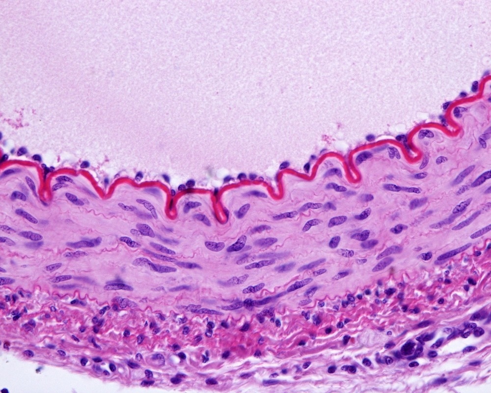
Study: Anionic nanoplastic exposure induces endothelial leakiness. Image Credit: Jose Luis Calvo/Shutterstock.com
Generally, plastic undergoes degradation naturally as well as artificially through artificial processes and is transformed into nanoplastics. Very few studies are available related to the biological fingerprints of nanoplastics.
In a recent Nature Communications study, researchers sought to address the aforementioned gap in research and revealed that the introduction of different nanoplastic forms, such as poly(methyl methacrylate) (PMMA) and anionic polystyrene (PS), causes damage to the vascular endothelial cadherin junctions.
The Effect of Nanoplastics on Humans
Microplastics and nanoplastics are formed due to the biological, physical, and chemical degradation of plastics that, subsequently, accumulate in landfills and water bodies. Plastic waste comes from various industrial sectors, research institutes, and by-products of different commodities (e.g., shampoos, and clothing). It is imperative to understand and formulate effective mitigating measures to deal with the adverse biological effects of nanoplastics in humans.
Different forms of nanoplastics are present in the air, soil, and water, and scientists have detected their traces in animals and human organs. These nanoplastics might have been introduced to humans and animals via inhalation, dermal exposure, and ingestion.
Nanoplastics affect humans in different ways; for example, they can impair growth, metabolism, or reproductive capacity. They also impact the immune system by enhancing endoplasmic reticulum stress, oxidative stress, cytokine secretion, and apoptosis.
Biological Fingerprints of Anionic Nanoparticles on Humans
The effect of anionic nanoparticles (less than 100 nm in size), such as titanium dioxide, gold, silicon dioxide, and nanodiamonds, is that it damages the vascular endothelial cadherin (VE-cadherin) junctions. They form a transient, small-sized physical opening in endothelial monolayers known as nanomaterial-induced endothelial leakiness (NanoEL). Nanotoxicological studies revealed NanoEL to exhibit a significant toxic effect.
Initially, PS nanoplastic was characterized using dynamic light scattering (DLS) and transmission electron microscopy (TEM). The average size of monodispersed PS beads was estimated to be approximately 21.2 nm in water and 26.2 nm in endothelial cell medium (ECM), via TEM analysis and around 62 nm in water and 72 nm in ECM, by the DLS method. The presence of carboxyls on nanoplastics was detected via X-ray photoelectron spectroscopy (XPS) analysis.
Nanotoxicology-based in vitro assays, namely, Cell Counting Kit-8 (CCK 8), reactive oxygen species (ROS), and cellular mortality assays, were conducted. In these assays, different concentrations of the nanoplastics were introduced to human umbilical vein endothelial cells (HUVECs) over time.
These experimental assays displayed impaired cellular membranes and DNA structures after twenty-two hours of treatment. After six hours of treatment, PS nanoplastics triggered autophagy and apoptosis by restricting the PI3K/AKT pathway in HUVECs. No cell death occurred up to ten hours of incubation with varied nanoplastic concentrations; however, cell shrinkage and deformities were observed. The amount of PS nanoplastics that penetrated HUVECs was increased over time and dosage.
Transwell assay with a fluorescent probe indicated that endothelial leakiness in HUVECs was triggered by the nanoplastics. Within one hour of the cells being exposed to 0.5 mg/mL of the nanoplastic, significant leakiness was observed. After six hours, all nanoplastic concentrations induced endothelial leakiness.
In addition to PS, NanoEL's competence in other nanoplastic forms, such as aminated polystyrene (NH2-PS) and PMMA, was studied. Both nanoplastics were characterized using varied analytical tools and were introduced to HUVECs.
After one hour of NH2-PS nanoplastic exposure to HUVECs, at 0.05 mg/mL, no change in cell viability was detected. However, over time, cell mortality was elevated significantly with the enhancement of nanoplastic dosage. Interestingly, in the case of PMMA nanoplastics, better innocuity and biocompatibility were observed.
The most interesting aspect of the new study is uncovering the function of nanoplastics (polymeric material) to elicit NanoEL. This observation has been reported for the first time. Additionally, the densities of different nanoplastics forms, i.e., PS (∼1.05 g/m3) and PMMA (∼1.18 g/m3) were found to be significantly lower than 1.72 g/m3, which is the threshold level for NanoEL-competent inorganic nanoparticles.
Two endocytosis inhibitors, namely, monodansylcadaverine (MDC) and methyl-β-cyclodextrin (MβCD), failed to stop the PS nanoplastic-induced leakiness; however, PP1 and Y-27632 were able to lower the pace of the process. Furthermore, several in vitro and in vivo assays strongly indicated that PS nanoplastic-induced endothelial leakiness was not dependent on endocytosis and ROS formation. Nevertheless, they were found to be strongly associated with actin remodeling and the VE-cadherin pathway.
To test if PS nanoplastics could induce leakiness in subcutaneous blood vessels, scientists administered Evans blue dye (EBD) into a mice's tail followed by either a control (phosphate buffered saline buffer) or PS nanoplastic. This experiment confirmed the incidence of endothelial leakiness by PS nanoplastic.
Future Outlook
The authors believe that the finding of the current study has opened up new avenues for studying the biological effects and behavior of different nanoplastics. This will positively help develop important guidance to build a sustainable plastics industry and many environmental remediation processes.
Reference
Wei, W., Li, Y., Lee, M. et al. (2022) Anionic nanoplastic exposure induces endothelial leakiness. Nature Communications, 13(4757). https://doi.org/10.1038/s41467-022-32532-5
Disclaimer: The views expressed here are those of the author expressed in their private capacity and do not necessarily represent the views of AZoM.com Limited T/A AZoNetwork the owner and operator of this website. This disclaimer forms part of the Terms and conditions of use of this website.