Germany’s Ruhr University Bochum and the Fraunhofer Institute for Microelectronic Circuits and Systems (IMS) have created a procedure that permits a novel type of signal amplification for diagnostic testing. Luminescent single-walled carbon nanotubes are being used in bioanalytics to perform more sensitive, economical, and timely tests. Enzymatic processes can be employed with the sensors.
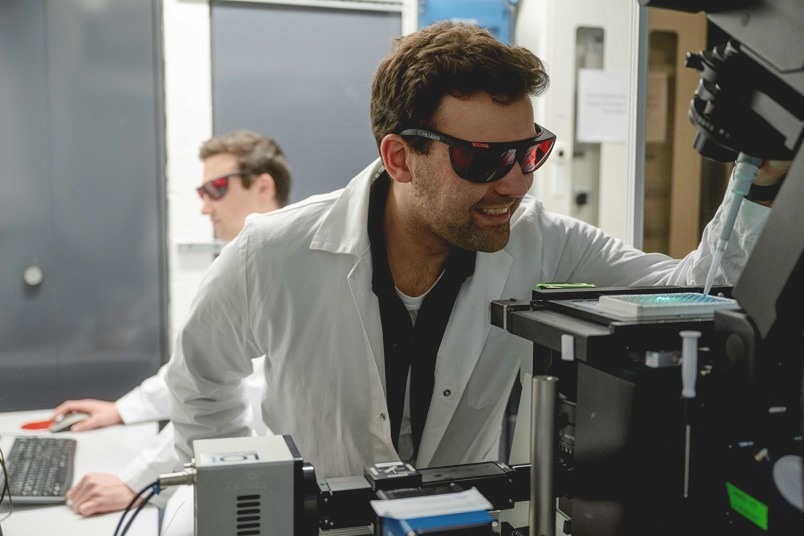
 Using fluorescent nanotubes, the researchers can detect the amount of certain substances. They use various optical setups to do this. Image Credit: RUB, Marquard
Enzyme-linked immunosorbent assays, or ELISAs for short, have a broad variety of applications due to their versatility in reaction conditions. The findings were released in the journal Angewandte Chemie International Edition on December 15, 2023. They provide fresh opportunities to enhance diagnostic processes and preserve detection agents.
Diagnostic Limits Can Be Improved with Luminous Carbon Sensor
Light is used in several diagnostic procedures to measure a substance’s concentration. This might be a luminescent or colorful substance. Unfortunately, the visible light range is full of background signals. Carbon tubes with a diameter of less than one nanometer were employed by the researchers to move the optical signal of measurement into a better spectral band. As compared to human hair, this is almost 100,000 times thinner.
The sensors fluoresce in the near-infrared spectrum, which is invisible to the naked eye, and they do not bleach. Furthermore, the sensors’ fluorescence is sensitive to their chemical environment as a result of a surface change. This allows for the observation and detection of chemical processes as they interact with the nanotube.
The fluorescence of the nanotubes pushes the signal into the near-infrared spectrum, which, combined with the nanotubes’ high sensitivity, causes a change in the detection threshold. This is significant, for example, when disease indicators are present at extremely low levels in an infection or disease like cancer.
Shifting the Detection Limit with Sensitive Nanosensors
The ability to customize nanotubes to different analytes offers a variety of possibilities, including increased sensitivity. This increase in sensitivity enables a possible change in detection limits, which can result in material and time savings in diagnostic operations. This new methodology has the potential to significantly enhance the efficiency of detection procedures in medical diagnostics.
Sensor Detects Different Substrates
The researchers proved that the novel sensor method works with the horseradish peroxidase enzyme’s substrates, p-phenylenediamine and tetramethylbenzidine.
This enzyme is used in a variety of biochemical detection methods. In principle, however, the concept can be applied to all kinds of systems. For example, we have also investigated the enzyme β-galactosidase, which is of interest for diagnostic applications. With a few modifications, it could also be used in bioreactions.
Justus Metternich, Ph.D. Student, Ruhr University Bochum
In the future, the group intends to modify the sensors for additional applications. For example, depending on the application, quantum flaws might be used to improve sensor stability.
This would be particularly advantageous if you not only want to measure in simple aqueous solutions, but also want to follow enzymatic reactions in complicated environments with cells, in the blood or in a bioreactor itself.
Sebastian Kruss, Professor of Physical Chemistry at Ruhr University Bochum
Kruss also heads the Fraunhofer IMS’s Attract Group Biomedical Nanosensors.
The study was supported by the Fraunhofer Attract Programme (038-610097), the German Research Foundation as part of the RESOLV Cluster of Excellence (EXC 2033-390677874), and the VW Foundation.
Journal Reference:
Metternich, J. T., et. al. (2024) Signal Amplification and Near-Infrared Translation of Enzymatic Reactions by Nanosensors. Angewandte Chemie International Edition. doi:10.1002/anie.202316965