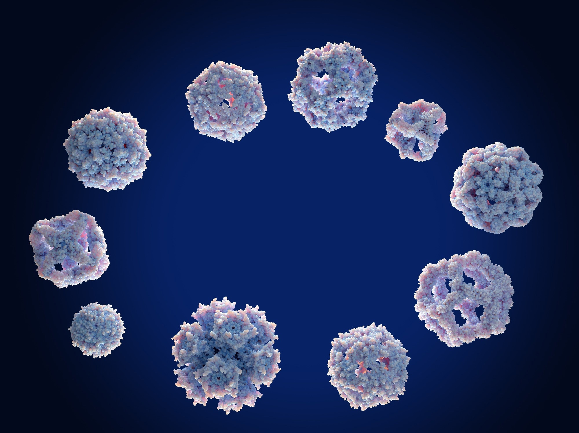
In a recent article published in Scientific Reports, researchers from Egypt developed new bimetallic nano-complexes to target specific proteins associated with breast and liver cancer. The research focuses on the potential of these complexes as therapeutic agents, highlighting their unique properties and the significance of their interactions with cancer-related proteins.

Image Credit: Juan Gaertner/Shutterstock.com
Background
Cancer remains a leading cause of mortality worldwide, necessitating the development of novel treatment strategies. This study focuses on targeting specific proteins, such as 3S7S in breast cancer and 4OO6 in liver cancer, to enhance therapeutic efficacy.
Previous research has shown that metal complexes can exhibit anticancer properties, prompting the exploration of bimetallic complexes in this study.
The Current Study
The bimetallic complexes were synthesized using a stepwise approach involving the reaction of metal ions (Mn(II), Ni(II), Cu(II), Zn(II), Hg(II), and Ag(I) with a multifunctional ether ligand. Comprehensive characterization was performed using Infrared (IR) Spectroscopy, Ultraviolet-Visible (UV-Vis) Spectroscopy, and Magnetic Resonance (NMR) Spectroscopy to determine their elemental composition, functional groups, coordination modes, and electronic transitions.
Mass spectrometry confirmed the molecular weight and identity of the complexes. Electron Spin Resonance (ESR) Spectroscopy investigated the electronic environment of unpaired electrons in the metal complexes.
Thermogravimetric analysis (TGA) and differential thermal analysis (DTA) assessed thermal stability and decomposition behavior, while magnetic susceptibility measurements provided insights into the electronic configurations of the metal ions. Scanning electron microscopy (SEM) and transmission electron microscopy (TEM) visualized the morphology and size of the nanoparticles.
The cytotoxic effects of the synthesized complexes were evaluated using in vitro assays against breast cancer cell lines expressing the 3S7S protein and liver cancer cell lines expressing the 4OO6 protein. Cell viability was assessed using the MTT assay, determining IC₅₀ values for each complex to evaluate their potency.
Results and Discussion
The elemental analysis results showed a match between the observed ratios of carbon, hydrogen, and nitrogen and the proposed stoichiometric molecular formulas. The IR spectra of the complexes exhibited the disappearance of the free –NH stretching vibration in the ligand spectrum, which suggested coordination through the Nitrogen atom to the metal center. Additionally, new bands that were attributed to metal-ligand bonding appeared, confirming the formation of coordination complexes.
The UV-Vis spectra revealed distinct absorption peaks indicative of d-d transitions within the metal ions. The position and intensity of these peaks varied with different metal ions, suggesting that the electronic environment around the metal centers was influenced by the ligand's coordination mode. The observed shifts in absorption maxima further supported the successful formation of the metal-ligand complexes.
The Proton-NMR spectra of the ligand and its metal complexes displayed significant chemical shift changes, particularly in the aromatic and aliphatic regions. These shifts were indicative of the ligand's interaction with the metal ions, confirming the coordination of the ligand to the metal centers.
Mass spectrometric analysis showed the presence of specific ion peaks corresponding to the metal-ligand complexes, further validating the successful synthesis. The TGA and DTA thermal profiles revealed multiple weight-loss steps attributed to the loss of coordinated solvent molecules and the breakdown of the ligand framework. This thermal stability is crucial for potential applications in drug delivery systems.
The magnetic susceptibility measurements indicated that the complexes exhibited paramagnetic behavior, characteristic of transition metal complexes. The SEM and TEM analyses revealed that the synthesized complexes formed spherical nanoparticles with size in the 50-100 nm range, which is favorable for cellular uptake in biological applications.
The results from the MTT assay demonstrated that the complexes exhibited significant dose-dependent cytotoxic effects against breast cancer (3S7S) and liver cancer (4OO6) cell lines. The calculated IC₅₀ values for the complexes indicated varying degrees of potency, with some complexes showing superior activity compared to the reference drug, Cisplatin.
Notably, the Cu(II) complex displayed the lowest IC₅₀ value, suggesting a strong potential for therapeutic application in cancer treatment. The observed cytotoxic effects were further supported by the hypothesis that the metal ions could interact with cellular components, generating reactive oxygen species (ROS). This oxidative stress can trigger apoptosis in cancer cells, thereby enhancing the therapeutic efficacy of the complexes.
Conclusion
The research concludes that the newly synthesized nano-complexes show promise for targeting cancer proteins, potentially leading to innovative breast and liver cancer treatment options. The study underscores the importance of further investigation into the biological activity of these complexes and their potential role in enhancing cancer treatment strategies.
Future research directions include exploring the therapeutic efficacy of these complexes in clinical settings.
Journal Reference
Faheem SM., et al. (2024). New nano-complexes targeting protein 3S7S in breast cancer and protein 4OO6 in liver cancer investigated in cell line. Scientific Reports. DOI: 10.1038/s41598-024-65775-x