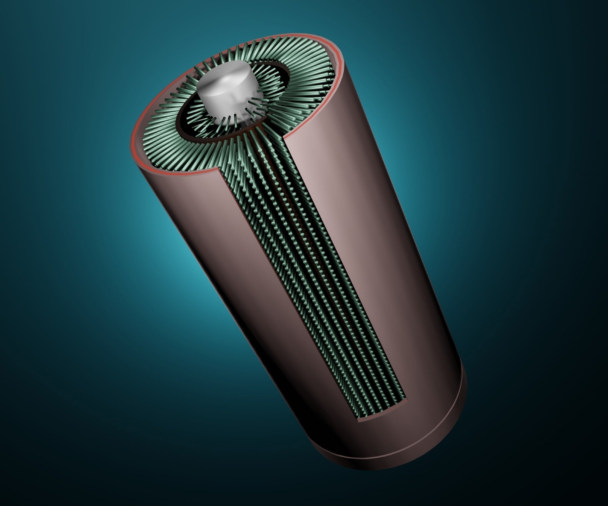
In a recent article published in Electrochemistry, researchers investigated the enhancement of extracellular electron transfer (EET) through nanowire (NW) electrode structures, focusing on the role of multi-heme cytochromes on the bacterial cell surface. The study aims to improve electron transfer efficiency by optimizing electrode design, thereby advancing the application of Desulfovibrio ferrophilus IS5 in bioelectrochemical systems.

Image Credit: Love Employee/Shutterstock.com
Background
The increasing demand for sustainable energy solutions has sparked interest in bioelectrochemical systems, particularly those that utilize microorganisms for energy conversion and storage. Desulfovibrio ferrophilus IS5, a sulfate-reducing bacterium, has gained attention for its ability to transfer electrons directly to electrodes—a process crucial for developing microbial fuel cells and bioremediation technologies.
Extracellular electron transfer involves the movement of electrons from microbial cells to solid electron acceptors, such as electrodes. In D. ferrophilus IS5, this process is facilitated by multi-heme cytochromes, integral membrane proteins that play a key role in the electron transport chain. Various factors, including electrode surface properties and the physical arrangement of bacterial cells, can influence the efficiency of EET.
The Current Study
Boron-doped p-Si(100) substrates were cleaned using ultrasonic baths in deionized water, acetone, and ethanol. A thin layer of gold (Au) was deposited via thermal evaporation to serve as a catalyst for metal-assisted etching. The substrates were then immersed in a solution of hydrogen peroxide (H2O2) and hydrofluoric acid (HF) to selectively etch the silicon, forming vertically aligned silicon nanowires of varying lengths (50 nm, 200 nm, and 500 nm). After etching, the nanowires were coated with a 20 nm layer of indium tin oxide (ITO) using sputtering to enhance electrical conductivity.
Desulfovibrio ferrophilus IS5 was cultured in modified Postgate's B medium with lactate as the electron donor and sulfate as the electron acceptor. The culture was incubated anaerobically at 30 °C, and cells were harvested during the exponential growth phase. The bacterial suspension was adjusted to an optical density of 0.5 at 600 nm in an anoxic electrolyte solution.
A three-electrode system was employed, with the NW electrode as the working electrode, a saturated calomel electrode as the reference, and a platinum wire as the counter electrode. Single-potential amperometry was performed at +0.4 V vs. SHE, while differential pulse voltammetry (DPV) was conducted with a 5.0 mV pulse increment and 50 mV pulse amplitude. Background currents were subtracted using QSoas software.
Current densities were calculated by normalizing currents to the electrode surface area. DPV peak currents were correlated with redox-active species concentrations, allowing for the estimation of electron transfer rates. Statistical analyses were performed to assess the significance of differences among the various NW configurations.
Results and Discussion
The study demonstrated that nanowire-arrayed electrodes significantly improved the rate of extracellular electron transfer compared to flat ITO electrodes.
Scanning electron microscopy (SEM) images revealed densely packed nanowire arrays, which enhanced the hydrophilicity of the electrode surfaces. This increased hydrophilicity is believed to promote better cell attachment and facilitate the electron transfer process.
The length of the nanowires was found to play a critical role in determining the efficiency of EET, with longer nanowires showing enhanced performance. The optimal length for maximizing electron transfer was identified, indicating that the spatial arrangement of the nanowires can be fine-tuned for the best results.
Electrochemical measurements showed that the nanowire electrodes exhibited higher current densities in the presence of D. ferrophilus IS5, supporting the hypothesis that nanostructured surfaces can enhance microbial electron transfer.
The study also emphasized the importance of multi-heme cytochromes in the electron transfer mechanism, as these proteins are essential for the direct transfer of electrons from bacterial cells to the electrode surface. The findings align with previous research, which has established the critical role of cytochromes in facilitating EET in various microorganisms.
Conclusion
This research provides valuable insights into enhancing extracellular electron transfer in Desulfovibrio ferrophilus IS5 using nanowire electrode structures.
The study successfully demonstrated that nanowire arrays significantly improve electron transfer efficiency, primarily due to their increased surface area and hydrophilicity, which promote better cell attachment and interaction with multi-heme cytochromes.
The findings underscore the potential of nanostructured electrodes in advancing bioelectrochemical systems, paving the way for more efficient microbial fuel cells and bioremediation technologies. Future research should focus on further optimizing nanowire electrode design and exploring the mechanisms underlying enhanced EET to fully harness microorganisms' capabilities in sustainable energy applications.
Journal Reference
Deng X.., et al. (2024). Nanowire Electrode Structures Enhanced Direct Extracellular Electron Transport via Cell-Surface Multi-Heme Cytochromes in Desulfovibrio ferrophilus IS5. Electrochemistry. DOI: 10.3390/electrochem5030021, https://www.mdpi.com/2673-3293/5/3/21