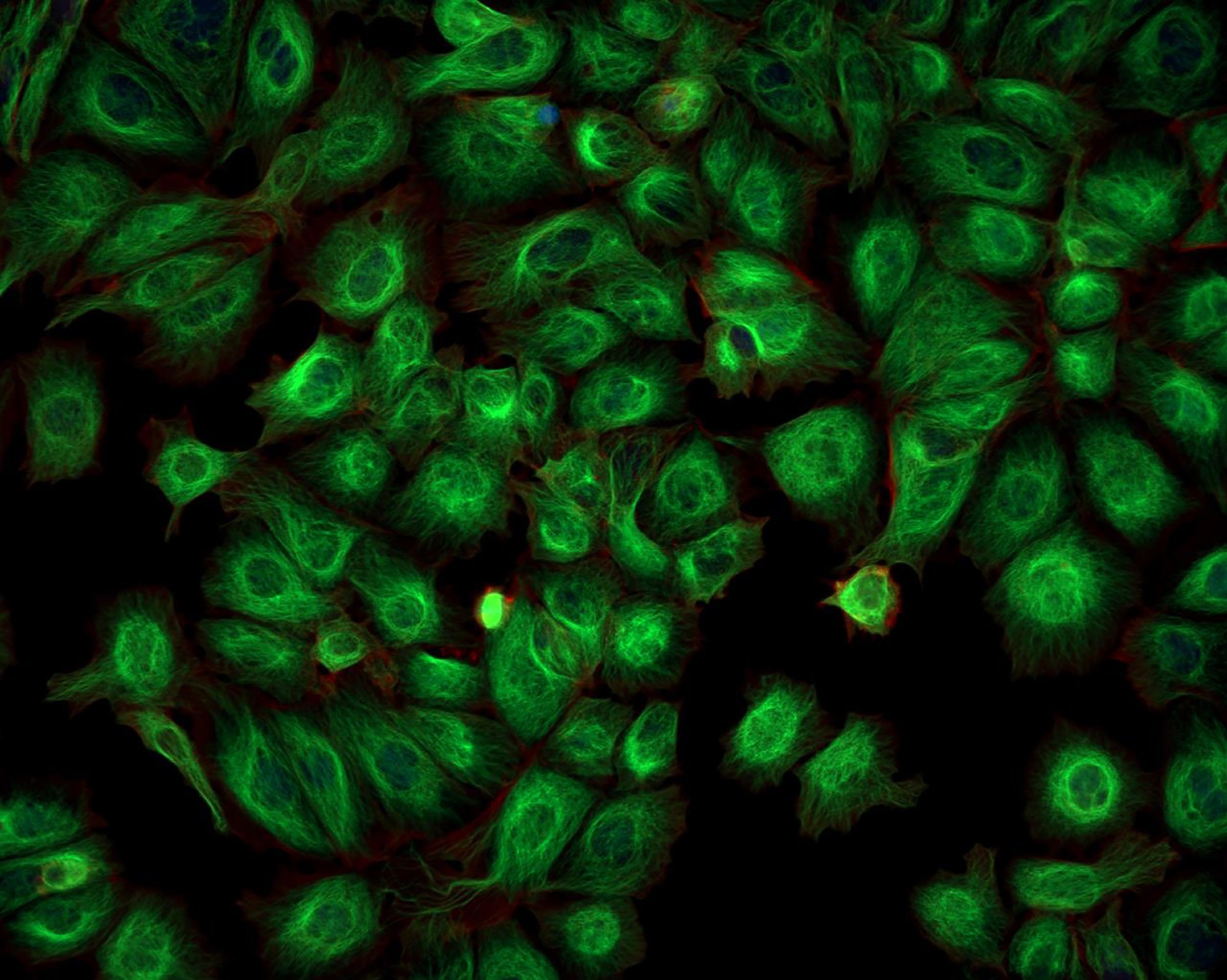
In a recent Nature Communications article, researchers introduced destabilized near-infrared (NIR) fluorescent nanobodies designed to target green fluorescent protein (GFP)-based biosensors. These nanobodies enable background-free imaging and manipulation of biological processes, offering improved specificity and sensitivity, particularly in live animal models.

Image Credit: Caleb Foster/Shutterstock.com
Background
The growing demand for precise imaging techniques in biological research has highlighted the limitations of traditional fluorescent proteins, especially in live animal models. While GFP and its derivatives are commonly used for cellular visualization, they often face issues like photobleaching, high background fluorescence, and limited tissue penetration. These challenges obscure the dynamics of cellular processes and hinder the study of complex biological interactions in real time.
Near-infrared (NIR) fluorescent proteins offer a promising alternative due to their ability to penetrate deeper into tissues and reduce interference from surrounding structures. However, developing NIR probes with high specificity and sensitivity remains a challenge. This study addresses this gap by introducing destabilized NIR fluorescent nanobodies designed for background-free imaging of GFP-based biosensors, improving the accuracy and reliability of in vivo imaging techniques.
The Current Study
The researchers used a series of molecular biology techniques to develop and characterize the NIR fluorescent nanobodies. They began by constructing plasmids encoding the nanobodies, which were then expressed in suitable host cells. After purification, the nanobodies were characterized for their binding affinity and fluorescence properties.
To test their functionality in vivo, the researchers injected diluted adeno-associated virus (AAV) containing GFP constructs into the somatosensory cortex of mice, following established surgical protocols to ensure accurate and safe injections.
After the injections, the mice underwent transcardial perfusion to preserve brain tissue for further analysis. Brain sections were prepared using a vibratome and immunostained with specific antibodies to visualize GFP expression and NIR nanobody binding. Confocal microscopy was then used to capture high-resolution images of the brain sections, enabling detailed analysis of the spatial distribution of the nanobodies and their interaction with the target proteins.
Results and Discussion
The results showed that the destabilized NIR fluorescent nanobodies successfully bound to GFP in both in vitro and in vivo settings.
The researchers observed a significant reduction in background fluorescence, greatly enhancing the clarity of images from live tissues. These imaging experiments demonstrated that NIR nanobodies could visualize cellular processes with high specificity, allowing for clear differentiation between GFP-expressing and non-expressing cells.
Additionally, the study highlighted the potential of these nanobodies for multiplexing, enabling the simultaneous visualization of multiple targets without interference.
The authors discussed the broader implications of their findings, noting that the ability to manipulate and visualize biological processes in real time opens up new opportunities for studying complex cellular interactions and signaling pathways. NIR fluorescent nanobodies could be particularly beneficial in optogenetics, where precise control of neuronal activity is crucial for understanding brain function.
The study also suggested that these nanobodies could be adapted for use with other fluorescent proteins, expanding their applicability across various research fields.
The researchers highlighted the need for further optimization of the nanobodies for specific applications. While the results are promising, they acknowledged that more experiments are needed to fully understand the dynamics of nanobody-target interactions in live systems. They also emphasized the importance of considering factors such as tissue heterogeneity and potential off-target effects when interpreting results.
Conclusion
The development of destabilized NIR fluorescent nanobodies marks a significant advancement in molecular imaging. This approach enhances the specificity and sensitivity of imaging techniques, enabling clearer visualization of biological processes in live animals.
The study provides a solid foundation for future research aimed at optimizing these nanobodies for various applications, including optogenetics and multiplexed imaging. By addressing the limitations of traditional fluorescent proteins, the authors have opened new avenues for exploring complex biological systems with unprecedented clarity and precision.
The potential applications of these nanobodies in both basic research and clinical settings underscore their importance in advancing our understanding of cellular dynamics and interactions. As the field of molecular imaging continues to evolve, the integration of NIR fluorescent nanobodies into existing methodologies promises to enhance our ability to study and manipulate biological processes in real time.
More from AZoNano: Ethics in Nanomedicine: Key Issues and Principles
Journal Reference
Barykina, N.V., et al. (2024). Destabilized near-infrared fluorescent nanobodies enable background-free targeting of GFP-based biosensors for imaging and manipulation. Nat Commun. https://doi.org/10.1038/s41467-024-51857-x, https://www.nature.com/articles/s41467-024-51857-x