Reviewed by Lexie CornerNov 7 2024
According to a study published in Science, researchers have created a new record-breaking 3D imaging tool. The tool employs high-energy X-rays to reveal or “visualize” the internal structure of nanomaterials.
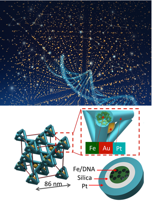
 Visualization of a 3D assembly of DNA-guided nanoparticles and frameworks (top). Multi-element template (teal) framework of tetrahedra (bottom). Zoomed-in portion shows iron (Fe), silica, platinum (Pt), and gold (Au). Image Credit: Columbia University and Brookhaven National Laboratory
3D nanostructures are critical to advancing many applications. Composed of nanomaterials—natural and synthetic substances that are more than 10,000 times thinner than a human hair—these structures are essential in a variety of fields, including the development of new biomaterials, catalysts, solar cells, and other devices that interact with light.
One method for creating these nanostructures involves using tailored DNA to program the assembly of the materials. This opens the door to new materials whose properties can be tuned at the nanoscale.
The Impact
To create nanostructures, researchers need to investigate their internal architecture in three dimensions at various stages of assembly. However, current nanoscale probing methods are limited. To generate X-ray computed tomography (CT) scans, the researchers employed a range of techniques, including powerful light sources.
These scans provided unprecedented resolution of 7 nanometers and detailed information about the elements within the materials. The researchers then constructed 3D frameworks to reveal the imperfections and interfaces of the nanostructures, which also clarified the assembly process of these structures.
Summary
Bottom-up nanofabrication is a promising approach for creating complex 3D materials by self-assembling functional nanocomponents. In particular, DNA encoding can provide a high level of structural and compositional control for programming unique and specific material organization.
In this study, the researchers developed a new self-assembling nanomaterial with novel catalytic, mechanical, and electronic properties. The research utilized the Center for Functional Nanomaterials (CFN), a DOE user facility at Brookhaven National Laboratory (BNL).
While this technique offered the researchers significant control over the designed material, it remained unclear how various designs and fabrication protocols influenced its structure and functionality.
The team developed an X-ray CT tool at the Hard X-ray Nanoprobe (HXN) beamline at NSLS-II, enabling the imaging of the inner architecture of nanomaterials. This was achieved by integrating the brilliant X-ray source of the National Synchrotron Light Source II (NSLS-II), another DOE user facility at Brookhaven National Laboratory (BNL), with nanofocusing multilayer Laue lenses.
A critical phase of this study involved creating new software tools to help break down the vast amounts of data into manageable chunks for processing and analysis. Through an iterative process, the team successfully visualized the 3D structures of individual nanoparticles and frameworks, overcoming the significant challenge of identifying various types of defects in the formed structures. They also confirmed the record-setting resolution of the X-ray microscopy using standard analysis and machine-learning techniques.
The synergistic capabilities of the new functional nanomaterials produced through bottom-up fabrication, combined with the sophisticated 3D X-ray CT tool and innovative analysis software, facilitate faster materials development and characterization.
The Department of Defense’s Army Research Office, the Department of Energy’s (DOE) Office of Science, and the Office of Basic Energy Sciences provided funding for this study. The Center for Functional Nanomaterials and the National Synchrotron Light Source II, both DOE Office of Science user facilities, supplied research resources.
Journal Reference:
Aaron, M. et. al. (2024) Three-dimensional visualization of nanoparticle lattices and multi-material frameworks. Science. doi.org/10.1126/science.abk0463