In this interview, Professor Emeritus Mervyn Miles at the University of Bristol speaks about the history and technology behind Atomic Force Microscopy (AFM) and Scanning Probe Microscopy (SPM).
Can you tell us more about your career and what excites you about SPM technologies?
I have had a passion for microscopy ever since I can remember. From age 10, I tried making an optical microscope with my Meccano set. During my PhD, I used a million-volt microscope, and in my postdoc, I used a two-kilovolt electron microscope. Eventually, I came to Scanning Tunneling Microscopy (STM). Microscopy has been a recurring theme throughout my life, and it is like I have been looking into that magical space you cannot see with your eyes.
Can you explain the basic principles behind AFM?
Atomic Force Microscopy (AFM) followed STM and involves measuring forces between a sharp tip and a sample surface, using mechanical interactions. Unlike STM, which measures quantum mechanical tunneling current, AFM is a totally mechanical system. It is almost like a Victorian instrument but with the modern technology needed for data recording. AFM leverages very localized measurements with sharp tips to get detailed images.
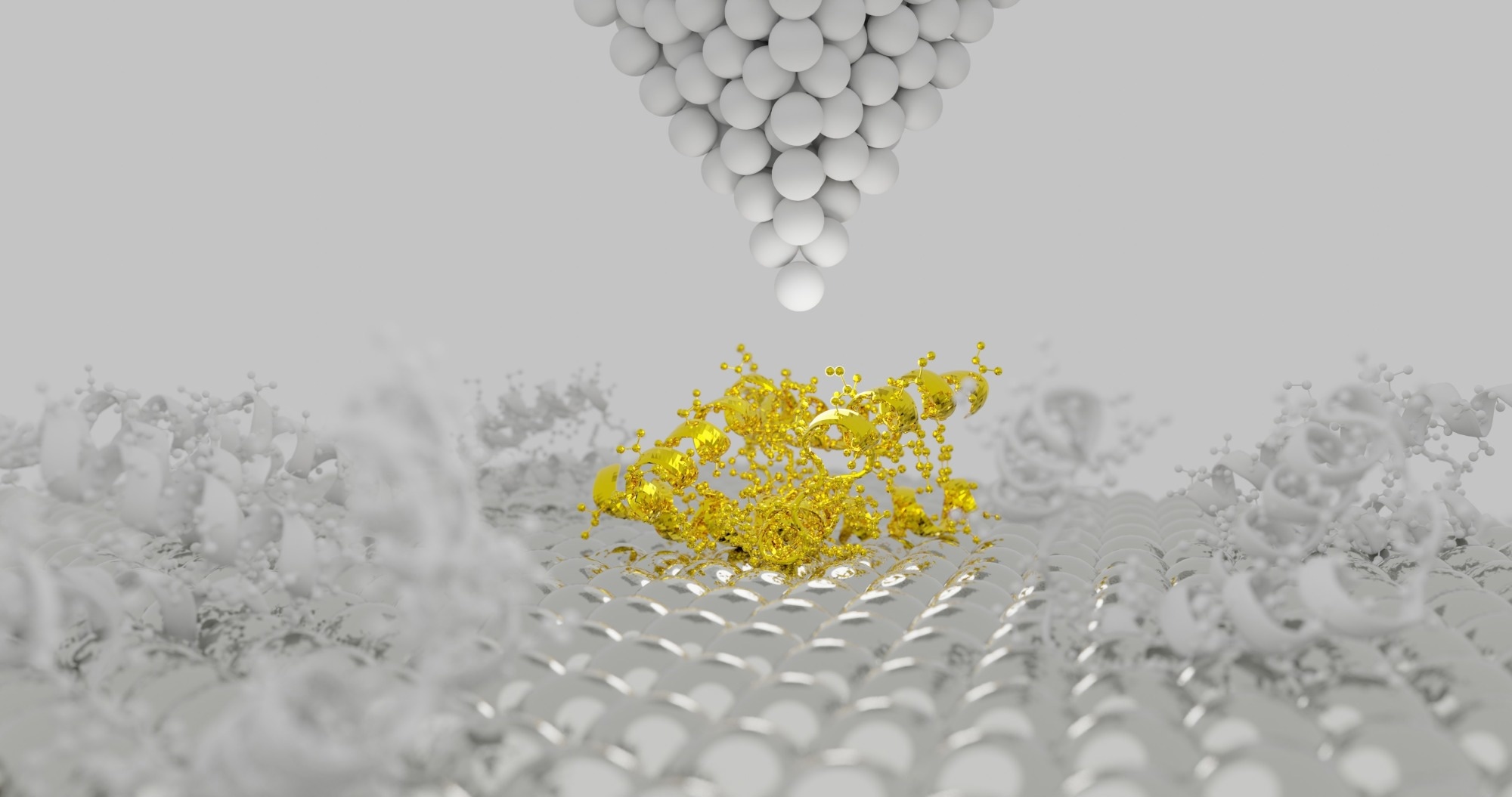
Image Credit: Shutterstock.com/Sanjaya Viraj Bandara
How does AFM overcome the resolution limits of optical microscopes, especially in life sciences?
AFM overcomes the resolution limits of optical microscopes by providing atomic resolution and working in liquid environments. Unlike optical microscopes, limited to about half a micron, AFM can achieve much finer resolution and give three-dimensional images in environments relevant to biological samples.
You met Heinrich Rohrer, who was the winner of the 1986 Nobel Prize in physics. How did that meeting impact your career?
I met Heinrich Rohrer at the first European Biophysics Congress in 1984. We walked together to the next session, and he explained STM to me. His explanation fascinated me. I realized the potential of STM and how revolutionary it could be. This meeting inspired me to pursue STM and AFM, significantly changing my career path.
Were proteins studied with STM and AFM from the beginning, or did research focus on other materials first?
In the early days of STM, the focus was on conductive samples. However, at the Institute of Food Research in Norwich, we explored biological molecules like proteins and polysaccharides. We were able to visualize individual protein molecules, which was amazing, even though the exact mechanism for imaging non-conductive samples was not fully understood.
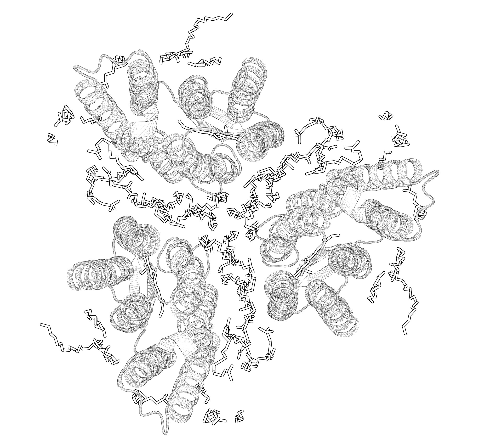
Image Credit: Shutterstock.com/Jean-Marc Billod
Did you switch completely to STM, or did you compare it with other methods like X-ray?
I did not completely switch from X-ray to STM but rather aimed to complement X-ray scattering data with direct imaging from STM. Combining these techniques allowed us to confirm models of molecular structures more accurately, making the research more reliable and comprehensive.
Are scientists still trying to combine different imaging methods with AFM, and how does that affect its use?
Integrating different imaging modes with AFM is crucial. For instance, combining AFM with optical microscopy for fluorescence measurements helps ensure that we are observing the correct part of the sample. This integration reduces bias and enhances the accuracy of our findings.
What were the early trends in AFM research, and what are the common applications now?
Initially, AFM research focused on structures like DNA, but recent trends emphasize high-speed imaging to capture real-time biological processes. Researchers like Toshio Ando have made significant advances, allowing us to follow dynamic biological processes, which is crucial for understanding how biological systems function.
Conversations on AFM #1: Interview with an AFM Pioneer
Video Credit: Bruker BioAFM
Do you think AFM will be used in medical applications soon, or is that still in the future?
Although AFM has great potential for medical diagnostics, its adoption has been slower due to the need for skilled operators. As AFM machines become more automated and user-friendly, I believe they will become more prevalent in the biomedical field, providing valuable insights into cellular mechanics and disease diagnostics.
What do you think is the future of AFM—automation of data collection or data analysis?
I see the future of AFM involving more automation in both data acquisition and analysis. I am particularly excited about vertical probe microscopy, which allows us to scan biological samples from various angles, providing truly three-dimensional images. Developing methods to look at samples from all directions will be a significant advancement, enabling us to study biological structures in their natural, three-dimensional contexts.
About Professor Emeritus Mervyn Miles
Mervyn Miles is a physicist renowned for pioneering innovative techniques in scanning-probe microscopy. This technology, which utilizes a nanometer-sized probe for surface scanning, has enabled unprecedented detailed visualization of biological systems, including molecules like lipids and DNA.
Among his significant contributions are methods for capturing over 100 images per second and techniques for probing delicate samples through a thin water layer, preventing contact damage. Additionally, he has enhanced image clarity and specimen preservation by immersing the microscope probe in liquid.
Currently, he is Professor Emeritus at the University of Bristol where he acted as head of the Nano Physics and Soft Matter Group. He held the prestigious position of Director of the Centre for Nanoscience and Quantum Information and was also a recipient of the Royal Society Wolfson Research Merit Award, Fellow of the Royal Society, and Honorary Fellow of the Royal Microscopical Society.

Click Here to Listen to the Podcast
This information has been sourced, reviewed and adapted from materials provided by Bruker BioAFM.
For more information on this source, please visit Bruker BioAFM.
Disclaimer: The views expressed here are those of the interviewee and do not necessarily represent the views of AZoM.com Limited (T/A) AZoNetwork, the owner and operator of this website. This disclaimer forms part of the Terms and Conditions of use of this website.