Quantum dots (QDs), semiconducting nanoparticles with 1-100 nm size, have emerged as advanced tools in biological research due to their unique optical and physical properties.1,2 These powerful tools allow researchers to explore intricate molecular interactions and cellular processes in living organisms in exceptional detail.1
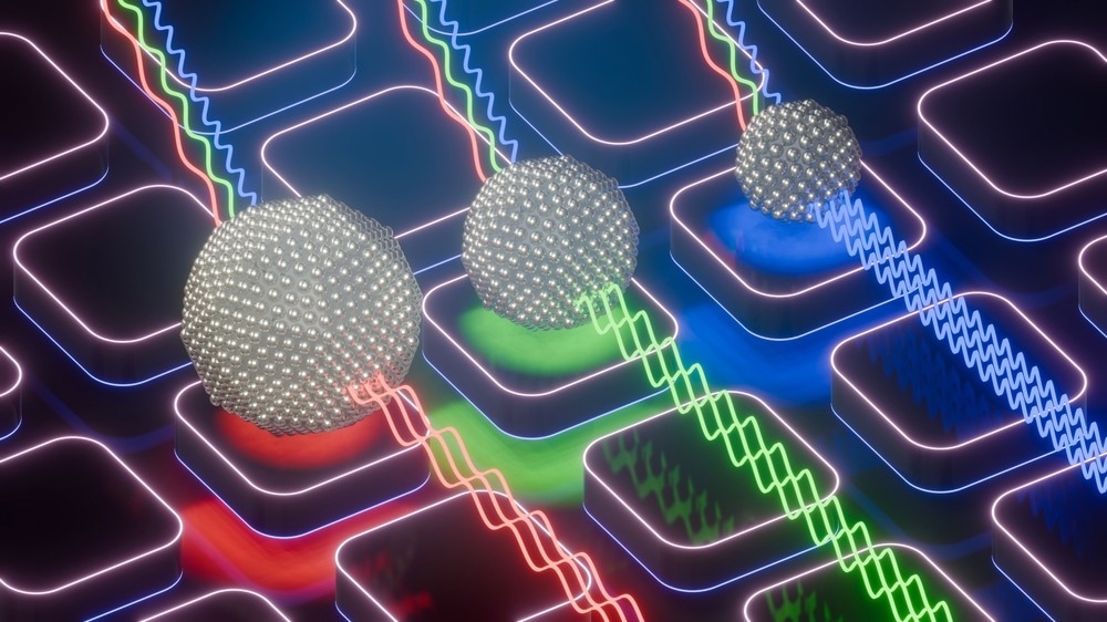
Image Credit: atdigit/Shutterstock.com
QDs offer excellent photobleaching-resistant fluorescence, making them highly suitable as fluorescence labels in biomedical imaging.2 Their size-dependent optical properties, such as exceptional photostability and quantum yield, help overcome the limitations of traditional fluorescent dyes.1
This article explores the mechanisms of QD labeling that enhance precision, sensitivity, and specificity in bioimaging applications.
Mechanisms of QD Labeling
QDs can bind covalently or non-covalently to biomolecules like antibodies, proteins, peptides, aptamer nucleic acids, small molecules, liposomes, lectins, and monosaccharides to identify specific targets. This binding provides QDs access to targets inside or outside a cell through various pathways, enabling their use in specific and non-specific targeting and biological imaging.1
Covalent binding involves reactions between QDs and functional groups on biomolecules, resulting in covalent binding of amino-carboxyl groups, amino-sulfhydryl groups, and aldehydes-hydrazides. This method ensures the selective binding of QDs to antibodies, enhancing specificity, stability, and water solubility.1
Non-covalent binding, on the other hand, can be hydrophobic or electrostatic between biomolecules and ligands on the QD surface. QDs are often encapsulated in silica or biocompatible polymeric shells, then combined with biomolecules through terminal groups on the shell, improving their biocompatibility and fluorescence stability.1
The photostability and brightness allow QDs to provide robust and reliable signals, even in challenging imaging conditions.1 For instance, QDs can reside intracellularly and label different hematological cell lines for extended periods. However, target ligands on QDs are essential for specific binding, otherwise they can be randomly internalized by multiple hematological cell lines.2
The physical attributes of QDs, including size, shape, and composition, allow tunability of their fluorescence across a broad spectrum. Researchers can tailor QD fluorescence by altering these parameters to suit specific imaging needs, which is highly advantageous in multiplexed imaging requiring differentiation between multiple targets.1
Applications in Research
QDs are versatile nanosized tools useful in various biological imaging applications, including single-molecule tracking, cell and tissue imaging, in vivo imaging, and multiplexed imaging. Their ability to provide insights into disease progression, treatment responses, and basic biological processes opens new possibilities in developmental biology, oncology, and neuroscience.1
QDs enable single-molecule tracking, allowing researchers to observe the dynamics of individual molecules and communication between living cells.1,2 For example, extracellular vesicles (EVs) secreted by cancer cells promote cancer’s proliferation and invasion. Therefore, QDs can be used as fluorescence probes to label and track the dynamics of EVs.2
A recent study in ACS Nano employed QDs as compact immunolabels for microtubule imaging and cell classification by conjugating them with specific antibodies. These QD-antibody conjugates allowed multispectral multiplexing through bright signals in the deep red and infrared and low steric hindrance. The proposed conjugates exhibited high functionality in cytological identification in fixed brain specimens.3
Another recent study in Cells demonstrated the interaction and biocompatibility of QDs with hematopoietic cells. Disorders such as myocardial infarction and stroke are associated with cardiovascular diseases (CVDs). Employing QDs in imaging cardiovascular systems helps determine the state of CVDs. Moreover, QDs can perform multiple functions such as carrying imaging, therapeutic, and targeting agents.4
Beyond whole-cell labeling, QDs are also used to label specific organelle structures. Due to their outstanding brightness from high quantum yield, QDs help understand the complex interactions of the immune system.2 The photostability of QDs also allows long-term imaging research, such as tracking viruses and probing real-time cellular processes.1,2
Advantages Over Traditional Dyes
Traditionally, organic dyes are used as fluoroprobes to visualize different cell structures. However, they possess limited efficiency in biological imaging due to short lifetimes and weak signal intensity.2
QDs, on the other hand, offer high photobleaching-resistant fluorescence (or photostability), allowing prolonged imaging sessions without signal degradation and providing consistent and high-quality images.1,2
The high quantum yield of QDs surpasses the capabilities of traditional fluorescent dyes, enabling enhanced resolution, stability, and specificity. Moreover, QDs have a lower risk of phototoxicity to biological samples, permitting safer long-term observations.1
Different functionalization methods allow the bioconjugation of QD surfaces with several biomolecules, such as antibodies, peptides, or nucleic acids. This allows specific targeting of biological structures or processes with high sensitivity and accuracy.
Moreover, biological coupling makes QDs multifunctional. They can be used for simultaneous imaging and treatment (such as photodynamic therapy) or combining different detection methods (such as fluorescence and magnetic resonance imaging).1
Multi-target detection without signal interference and low background noise to produce stable and reproducible results in different biological environments is another advantage of QDs over traditional dyes.1
Future Outlook and Challenges
Despite being powerful tools for biological research, QDs face multiple challenges that hinder their practical applicability. Their high toxicity and limited biocompatibility are primary concerns.1
The in vivo applications of QDs are limited by their potential toxicity. Strategies to minimize toxicity include adding ligands for structural stability and solubility and developing non-heavy-metal-containing QDs. However, these methods only partially mitigate QD toxicity.2
QD accumulation has been observed in the heart, lungs, kidneys, liver, and brain. It can trigger unwanted immune system responses. However, specific interactions between QDs and these organs at the tissue level are not well understood. Moreover, the long-term impact of this interaction and QD accumulation is unknown.
QDs also suffer from target-specificity issues. Modifying their ligands can increase target specificity, but this strategy can be problematic for in vivo applications, and minor off-target effects persist.2
With rising awareness of human-induced environmental damage, the biodegradability of QDs is a concern. Traditional QDs composed of heavy metals are non-biodegradable, and their accumulation in land and aquatic animals raises health concerns for the entire food chain. Efforts are being made to develop biodegradable QDs and greener synthesis methods, which can reduce environmental damage by recycling biowaste.2
Overall, the unique material constitution and structural design of QDs provide significant advantages over traditional fluorescent dyes, making them critical tools for advancing biological research.
Ongoing interdisciplinary technological developments, combining nanotechnology and biomedical knowledge, will help address current challenges, improving the safety and applicability of QDs as efficient medical imaging tools in the future.1
More from AZoM: Bio-based Polyethylene: A Sustainable Solution for Plastic Waste
References and Further Reading
1. Huang, S., Huang, G. (2024). The utilization of quantum dot labeling as a burgeoning technique in the field of biological imaging. RSC Advances. DOI: 10.1039/D4RA04402A
2. Le, N., Kim, K. (2023). Current Advances in the Biomedical Applications of Quantum Dots: Promises and Challenges. International Journal of Molecular Sciences. DOI: 10.3390/ijms241612682
3. Kolossov, V. L. et al. (2024). Quantum Dot-Fab′ Conjugates as Compact Immunolabels for Microtubule Imaging and Cell Classification. ACS Nano. DOI: 10.1021/acsnano.4c02215
4. Naylor-Adamson, L., Price, TW., Booth, Z., Stasiuk, GJ., Calaminus, SDJ. (2024). Quantum Dot Imaging Agents: Haematopoietic Cell Interactions and Biocompatibility. Cells. DOI: 10.3390/cells1304035
Disclaimer: The views expressed here are those of the author expressed in their private capacity and do not necessarily represent the views of AZoM.com Limited T/A AZoNetwork the owner and operator of this website. This disclaimer forms part of the Terms and conditions of use of this website.