A Brown University-led research team has made use of gold nanoparticles coated with a charged polymer ring and an X-ray scatter imaging technology to detect tumor-like masses as small as 5 mm.
The American Chemical Society’s Nano Letters journal reported the method in detail. In this method, metal nanoparticles are used as agents to improve X-ray scattering signals to identify tumor-like masses.
 A new diagnostic technique can spot tumor-like masses as small as 5 millimeters in the liver.
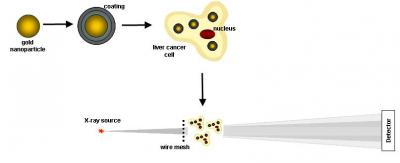
The research team selected gold nanoparticles with a diameter range of 10-50 nm and coated them with two layers of 1-nm polyelectrolyte rings. The coating offered the nanoparticles a charge that caused the nanoparticles to be engulfed by the cancerous cells. After being engulfed, the researchers utilized X-ray scatter imaging to identify the gold nanoparticles inside the affected cells.
In laboratory studies, although the volume of nontoxic gold nanoparticles used was only 0.0006% of the volume of the cell, yet they had the critical mass needed for identification by the X-ray scatter imaging equipment.
The researchers will now use the method for clinical study commencing this summer. They will load a cancer-targeting antibody into the nanoparticle carrier to detect liver tumors in mice. Jack Wands, who serves as professor of medical science at Brown University’s Warren Alpert Medical School and director of Rhode Island Hospital’s Liver Research Center, developed the antibody.
Wands stated that the researchers intend to combine gold nanoparticles and the antibody to spot the development of early-stage liver tumors using X-ray imaging. According to the scientists, the X-ray scatter imaging method could be utilized to identify the assemblies of nanoparticles in other organs.