A recent study published in the journal Nano Letters examines graphene's viability as a conducting neural surface capable of promoting cellular adhesion, nerve branching, and expansion.

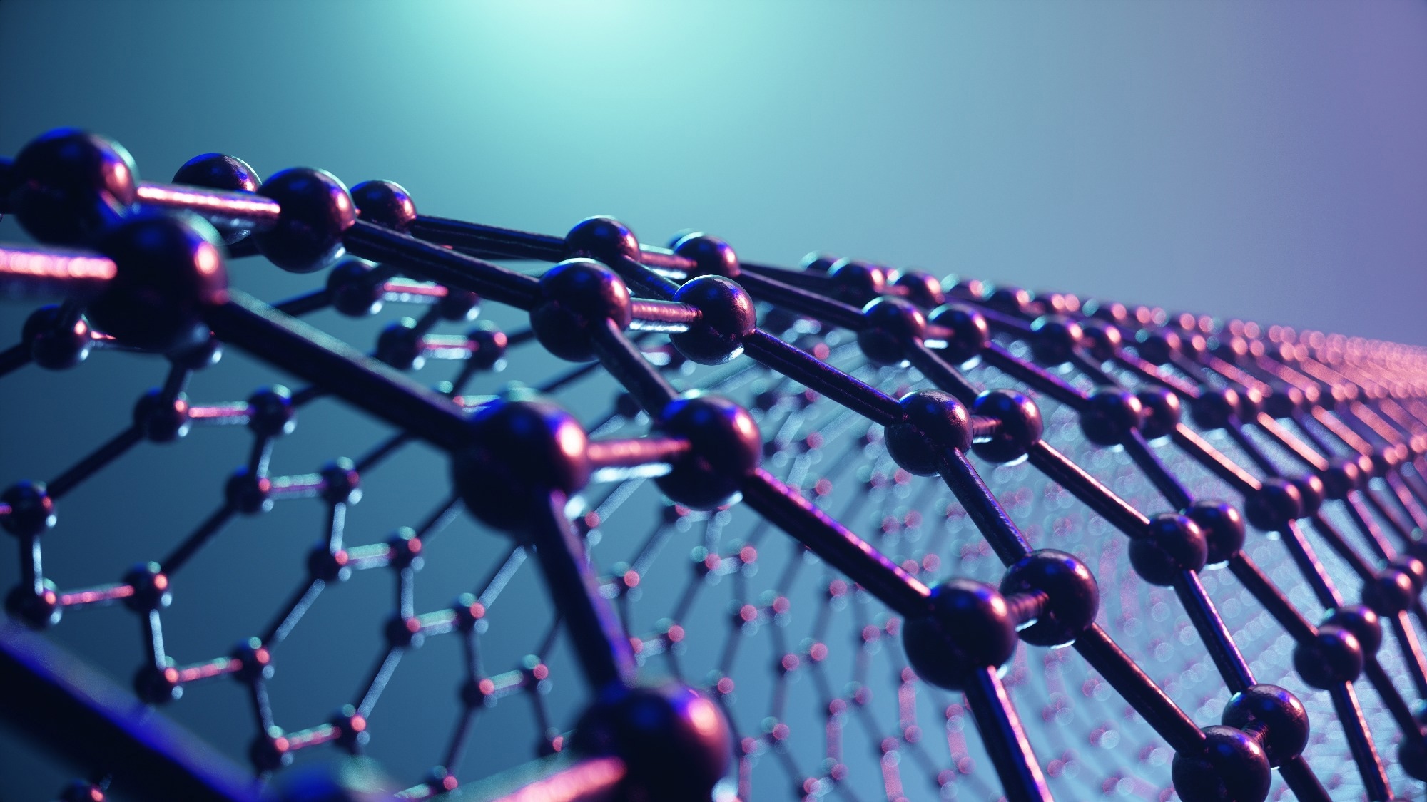
Study: Modulation of Early Stage Neuronal Outgrowth through Out-of-Plane Graphene. Image Credit: Rost9/Shutterstock
Neurons must incorporate a vast array of stimuli from their external surroundings for the proper construction of a neural network. Due to its astounding effectiveness, this mechanism plays a crucial part in the proper wiring of the cortex during growth and axonal regeneration. In this context, it has been shown that biologically motivated microstructured and nanostructured substrates, such as graphene, govern this axonal expansion.
Neural Tissue Engineering: Overview and Significance
The sophisticated neural circuitry that makes up the brain is formed by the elongation of axonal strands guided to their destinations by complicated navigation mechanisms. A system like this is required for the preliminary wiring of neuronal circuitry during brain growth and the rewiring of regenerated axons, which permits functional regeneration of brain circuitry after damage or illness.
Neural growth cones (GCs), sensory-motile elements positioned at the terminus of a stretching axon, control the inherent complexities of neuron orientation and neuronal network creation. GCs use a variety of chemical and geographical stimuli in their extracellular matrices to determine axon development and turning responsiveness.
A fundamental problem in neural tissue engineering is the designing of biologically inspired platforms capable of promoting and controlling axonal development through contact guiding mechanisms at the cell-material interface. For instance, anisotropic topographies, fissures, or fibers may be utilized to regulate axon orientation. Interrupted structures, such as nanowires or pillars, may also speed up neurite and axon development.
Graphene: The Future of Neural Tissue Engineering
Graphene is a carbon allotrope made from a single layer of atoms organized in a two-dimensional honeycomb lattice nanostructure. Graphene has become a desirable and versatile nanomaterial because of its outstanding mechanical strength, electrical conductance, opacity, and thinness.
Graphene's exceptional mix of features makes it an intriguing material platform for creating next-generation solutions in various fields, including ultrasensitive detectors, multidimensional materials, medicine, microbiology, and energy storage.
Graphene has recently piqued the attention of researchers in neurology and neural tissue engineering due to its unique chemical makeup, which includes a tremendously strong carbon bond, cytocompatibility, and high electrical conductance. The ability of graphene-based surfaces to induce neurite budding and outgrowth makes them great contenders for neural interfaces.
Although 2D graphene cultural systems are useful in studying the effect of graphene-based substrates on complicated cellular processes, new techniques aimed at imitating brain tissue intricacy in vitro are needed to control early-stage neuronal expansion.
What did the Researchers do in the Current Study?
In this study, the researchers showed the cell-instructive capability of micrometric out-of-plane graphene nanostructures. During the experiment, a three-dimensional fuzzy graphene structure (3DFG) was created on a collapsed silicon nanowire (SiNW) mesh template.
During the initial developmental phase of a neural network, optical imaging and the concentrated ion beam/scanning electron microscopy approach (FIB/SEM) were utilized to assess the cytocompatibility of the as-prepared graphene structures and their influence on growth cones (GCs) geometry and size. The cytoskeletal arrangement of GCs, primarily defined by actin and microtubule formations, was also studied.
The impact of out-of-plane graphene topographies inside GCs was analyzed and compared to the instance of two-dimensional graphene. Moreover, the neuron-to-graphene connection was studied from many perspectives, including integrin-mediated interface adhesion sites and cellular membranes curvature dynamics.
Key Developments of the Research
The as-produced 3D graphene structures were shown to be highly biocompatible with cortical neurons, allowing them to adhere and grow. The researchers demonstrated that graphene topographic structure and the micrometer characteristics of SiNW meshes impact growth cone (GC) formation and pathfinding activities, controlling GC shape, axon extension, bifurcation, and adherence.
Axons generated under the impact of 3D graphene surfaces were also longer and had fewer intermediate neurites. The micrometric protrusion characteristics of the nanowire meshes stimulated GCs to adopt a tiny bullet-shaped structure, a GC morphology more often observed in neurons with high navigational activity.
These results highlight the significance of developing nano-topographies for properly adjusting neuronal function at the cell-material interface. This study may also result in the invention of more efficient regenerative medicines since it provides a deeper knowledge of substrate-mediated effects on GC creation and axonal pathfinding activity.
Reference
Matino, L. et al. (2022). Modulation of Early Stage Neuronal Outgrowth through Out-of-Plane Graphene. Nano Letters. doi: https://pubs.acs.org/doi/10.1021/acs.nanolett.2c03171
Disclaimer: The views expressed here are those of the author expressed in their private capacity and do not necessarily represent the views of AZoM.com Limited T/A AZoNetwork the owner and operator of this website. This disclaimer forms part of the Terms and conditions of use of this website.