A recent study published in Nature Nanotechnology describes how urea-powered nanorobots helped a research team shrink the growth of bladder tumors in mice by 90%.
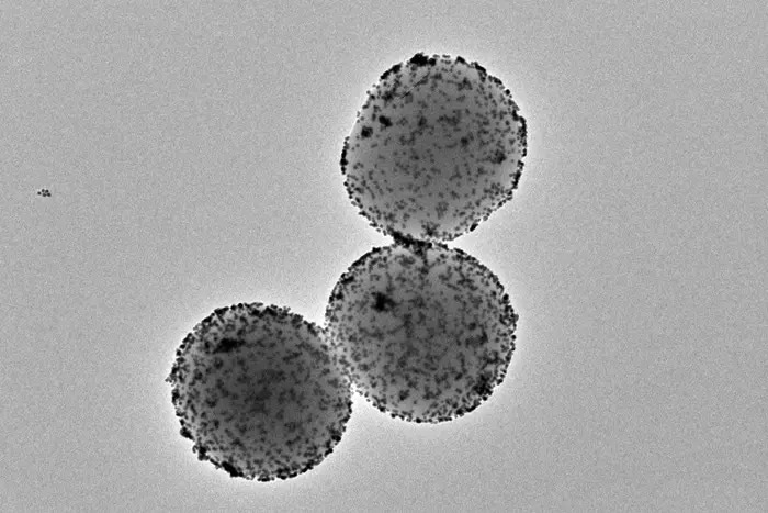
 Transmission electron microscopy image of the nanorobots. Image Credit: Institute for Bioengineering of Catalonia
Bladder cancer is the fourth most prevalent tumor in men and has one of the highest incidence rates in the whole world. Although bladder tumors have a low death rate, almost half of them recur within five years, necessitating continuous patient observation. This particular type of cancer is among the most expensive to treat due to recurrent hospital stays and the requirement for additional therapies.
Even while the current treatments that involve injecting drugs directly into the bladder have high survival rates, they nevertheless have limited therapeutic effectiveness. A possible substitute is using nanoparticles to deliver therapeutic drugs directly to the tumor. Among the most remarkable are nanorobots, which are nanoparticles that have the capacity to propel themselves within the body. These minuscule nanomachines are comprised of a permeable silica sphere.
On their surfaces are several components, each serving a distinct purpose. One is the urease enzyme, a protein that combines with urine’s urea to allow the nanoparticle to proliferate. Radioactive iodine, a radioisotope frequently employed for the localized therapy of tumors, is another essential element.
Innovative treatments for bladder cancer are made possible by this study, which is being directed by the Institute for Bioengineering of Catalonia (IBEC) and CIC biomaGUNE in conjunction with the Institute for Research in Biomedicine (IRB Barcelona) and the Autonomous University of Barcelona (UAB). The goal of these developments is to shorten hospital stays, which should result in cheaper expenses and more patient comfort.
With a single dose, we observed a 90% decrease in tumor volume. This is significantly more efficient given that that patients with this type of tumor typically have 6 to 14 hospital appointments with current treatments. Such a treatment approach would enhance efficiency, reducing the length of hospitalization and treatment costs.
Samuel Sánchez, Study Lead and ICREA Research Professor, Institute for Bioengineering of Catalonia
The investigation of whether these tumors reoccur after therapy is the next, and it is now under process.
A Fantastic Voyage into The Bladder
Scientists have previously demonstrated that nanorobots’ ability to propel themselves enables them to reach every bladder wall. This characteristic is favorable compared to the existing approach where, after injecting medication directly into the bladder, the patient must change position every half hour to ensure that the drug reaches all the walls.
This current study goes one step further by showing that nanoparticles are mobile inside the bladder and specifically accumulate in the tumor. This accomplishment was made possible by several methods, such as microscopy images of the tissues removed after the study was over and medical positron emission tomography (PET) imaging of the mice.
A fluorescence microscopy system created especially for this purpose at IRB Barcelona was used to capture the latter. By scanning the bladder’s many layers and producing a three-dimensional reconstruction, the technique makes it possible to view the organ in its entirety.
The innovative optical system that we have developed enabled us to eliminate the light reflected by the tumor itself, allowing us to identify and locate nanoparticles throughout the organ without prior labeling, at an unprecedented resolution. We observed that the nanorobots not only reached the tumor but also entered it, thereby enhancing the action of the radiopharmaceutical.
Julien Colombelli, Leader, Advanced Digital Microscopy Platform, IRB Barcelona
It was difficult to understand why nanorobots might infiltrate the tumor. Tumor tissue is generally stiffer than healthy tissue, and nanorobots lack the specialized antibodies to recognize the tumor.
However, we observed that these nanorobots can break down the extracellular matrix of the tumor by locally increasing the pH through a self-propelling chemical reaction. This phenomenon favored greater tumor penetration and was beneficial in achieving preferential accumulation in the tumor.
Meritxell Serra Casablancas, Study Co-First Author and Researcher, Institute for Bioengineering of Catalonia
The scientists deduced that although the urothelium crashes with the nanorobots as though it were a wall, the more porous tumor allows the nanorobots to pass through and gather within. The nanobots’ mobility is a crucial component that raises their chances of getting to the tumor.
According to Jordi Llop, a researcher at CIC biomaGUNE and co-leader of the study, “The localized administration of the nanorobots carrying the radioisotope reduces the probability of generating adverse effects, and the high accumulation in the tumor tissue favors the radiotherapeutic effect.”
“The results of this study open the door to the use of other radioisotopes with a greater capacity to induce therapeutic effects but whose use is restricted when administered systemically,” added Cristina Simó, co-first author of the study.
Years of Work and a Spin-Off
Results of more than three years of joint work across several universities are compiled in this study. A portion of the information is derived from the Ph.D. theses of Ana Hortelao and Meritxell Serra, two researchers working in Sánchez’s Smart nano-bio-devices department at IBEC. It also includes the thesis of Cristina Simó, co-first author of the study, who did her predoctoral research at the Radiochemistry and Nuclear Imaging Lab supervised by Jordi Llop at CIC biomaGUNE.
Another contribution is the animal model of the illness expertise of Esther Julián’s group at the UAB. Additionally, the “la Caixa” Foundation and the European Research Council (ERC) have provided financing for the study.
The foundation for Nanobots Therapeutics, a spin-off of IBEC and ICREA founded in January 2023, is the technology that has been in development for these nanorobots for more than seven years, thanks to Samuel Sánchez and his team’s recent patent.
Sánchez developed the company, which serves as a conduit for clinical application and research.
He added, “Securing robust funding for the spin-off is crucial to continue advancing this technology and, if all goes well, bring it to market and society. In June, just 5 months after the creation of Nanobots Tx, we successfully closed the first round of funding, and we are enthusiastic about the future.”
Technological Innovation in Microscopy to Locate Nanorobots
Working with nanorobots has presented a substantial scientific challenge in bioimaging approaches for detecting these components in tissues and the tumor. Common non-invasive clinical procedures, such as PET, lack the resolution to find these minute particles.
As a result, the Scientific Microscopy Platform at IRB Barcelona used a microscopy approach that illuminated materials with a sheet of laser light, allowing the capture of 3D pictures by light scattering upon interaction with tissues and particles.
The scientists created a novel approach based on polarized light that wipes out all scattering from the tumor tissue and cells after seeing that the tumor itself scattered some of the light, causing interference. This breakthrough allows for the imaging and placement of nanorobots without the requirement for previous molecular labeling.
Journal Reference:
Simo, C., et. al. (2023) Urease-powered nanobots for radionuclide bladder cancer therapy. Nature Nanotechnology. doi:10.1038/s41565-023-01577-y.