For the ninth year, JPK Instruments hosted the ninth annual international symposium on the applications of scanning probe microscopy (SPM) and optical tweezers.
Held in the historic Umspannwerk Ost in Berlin in early October, the meeting focused on applications developments in life sciences once more attracting over 100 scientists from around the world. The papers and parallel poster sessions inspired excellent discussions as attendees presented their results and shared scientific knowledge in a very relaxed and informal atmosphere.
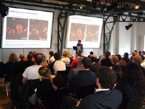
 The audience listen to Dr Claudio Canale at JPK's 2010 Life Sciences symposium
There were eighteen invited talks from leading scientists from Europe and Canada together with more than forty poster contributions provided a comprehensive overview about the current research topics in the fields of life sciences.
The SPM papers included several where proteins and biofilms were the subject of investigation. Scientists appear to have moved away from simply using the microscope to generate a surface picture but now want to get into the mechanics of how a film or cell may react under different conditions and probe forces. Morphology and mechanical changes are going a long way to show functionality so the SPM is really becoming a biophysical tool. Better calibration, more stable electronics enabling better system control and reproducibly-made probes means it is now possible to compare behaviour on the sub-pico Newton force scale. Cell adhesion measurements now have been given more relevance through simultaneous overlay of fluorescence imaging. An example was shown in the work of Dr Franz from Karlsruhe who clearly showed details of focal adhesions with total internal reflection fluorescence (TIRF) and their relevance to cellular adhesion. Dr Canale from Genova extended the study of fibrillar aggregates which provides information on early stage cytotoxicity.
The SPM poster award went to Arpita Roychoudhury of the University of Düsseldorf for her work on the stabilisation of membrane proteins using compatible solutes studied by force spectroscopy. This is looking at behavior at the molecular level. The goal of this study is designed to help in drug development for liver disease.
Day two saw the presentation of papers and posters applying optical tweezers technology. The technique is just beginning to show its full range of possibilities, in particular to measure and understand molecular forces and to study internal cellular changes. Users are moving towards using commercial solutions like Dr Modesti and his group at CNRS in Marseilles where he has a JPK NanoTracker system and studies the dynamics of nucleoprotein filaments which promote DNA sequence homology recognition and strand exchange. Homologous recombination is a universal life process, essential for maintenance of genome integrity, cell survival and protection against tumorigenesis.
The Optical Tweezers poster award was a challenge for the judges. There was a pull between applications of the technique and the art of instrumentation design. The 2010 winner was Anita Jannasch from the BIOTEC-TU Dresden. She presented single molecule Kinesin-8 measurements. Optical tweezers were used as both a force measurement and positioning tool. The work had fundamental value as it showed that kinesin could be attached to a microsphere without affecting its functionality.
As moderator of this year's meeting, Torsten Jähnke, Chief Technical Officer of JPK, said "the enthusiasm of delegates continues to surprise me particularly the ingenuity of those pushing applications to new levels. Sharing ideas to test their viability is so useful in the world where it is the scientist's wishes that drive the design for new instrumentation."
To learn more, visit the NanoBioVIEWS™ web site, www.nanobioviews.net. The abstracts of both papers and posters are now available to study online or may be downloaded in PDF format.