Although the advancement of microfluidic technologies accelerated the assay of circulating tumor cells (CTC), the release of the captured viable cells is still a challenge.

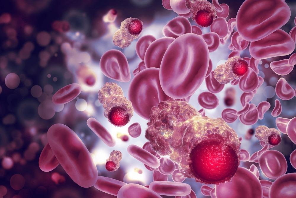
Study: pH-Responsive Carbon Nanotube Film-Based Microfluidic Chip for Efficient Capture and Release of Cancer Cells. Image Credit: crystal light/Shutterstock.com
In an article recently published in the journal ACS Applied Nano Materials, the authors fabricated a two component-based glass/polydimethylsiloxane (PDMS) microfluidic pH-responsive carbon nanotube (CNT) chip that can efficiently capture or release cancer cells in a patient blood sample.
CTCs and Their Release Strategies
CTCs shed from metastatic lesions, or primary tumors circulate in the bloodstream. They can be potentially used in liquid biopsies to detect cancer, help obtain genetic information on primary tumors, and choose a suitable therapeutic target.
Microfluidic technologies are convenient tools developed for CTC isolation from the bloodstream through their capture process that can be either a physical technique that exploits the biophysical properties of CTCs or an affinity-based method that involves immunocapturing of CTCs via CTC-specific antigens and release process.
However, despite the high selectivity of affinity-based approach toward CTCs, their release from the device for subsequent profiling is challenging. Although the inclusion of smart materials into immunocapture devices facilitated CTC release, these processes required lower temperatures or electric stimulation, which are detrimental to cells and incorporating photosensitive substrates to release CTCs caused DNA damage in the cells. Thus, there exists an urge for a robust release strategy that simultaneously ensures cell viability.
pH-Responsive CNT Film-Based Microfluidic Chip
In the present study, the authors fabricated a pH-responsive CNT film-based microfluidic chip to efficiently capture and release the epithelial cell adhesion molecule (EpCAM)-positive CTCs complex, useful for clinical evaluation and therapeutic prediction in the field of oncology.
The chip consisted of a bottom CNT-coated glass slide, functionalized with a pH-responsive poly-L-lysine (PLL) connected to EpCAM and a PDMS cover with herringbone microchannels on the top.
The PLL-connected anti-EpCAM antibodies capture CTCs. Herringbone microchannels on PDMS further enhance the capturing process. A subsequent change in pH can realize the release of CTCs with no effect on cells. This one-time-use device is biocompatible, have plenty of amino acids, and has a high specific area.
CNT Chip Characterization and Performance Evaluation
Scanning electron microscope (SEM) images of the as-prepared CNT film showed a spiderweb-like porous network of CNTs. Fourier transform infrared (FTIR) spectra of biotin-PLL showed characteristic peaks at 1547 and 1079-centimeter inverse, corresponding to amide and cyanide (CN) stretching. The peaks at 2923 and 2853-centimeter inverse correspond to asymmetric and symmetric stretching of methyl (CH2) in CNT. Furthermore, the peaks at 1731 and 1655-centimeter inverse correspond to carbonyl (C=O) stretching of the carboxyl group and alkene (C=C) stretching, respectively.
Raman spectra of the CNT film revealed a radial breathing mode (RBM), suggesting the presence of single-walled nanotubes. Furthermore, an upshift in RBM bands with biotin-PLL addition was due to a decrease in electron density. The lower intensities of RBM and G bands of biotin-PLL-CNT compared to CNT suggest the presence of cation−π interaction between the amine group of biotin-PLL and π-electrons of CNT.
The ultraviolet-visible (UV-vis) spectroscopy conducted on biotin-PLL−CNT and the spectra revealed the presence of two absorption peaks at 195.5 and 269 nanometers which are also present in pure biotin-PLL and CNT spectra. The bathochromic shift from 265.5 to 269 nanometers showed conjugation between biotin-PLL’s amino groups and π electrons.
CD spectroscopy studies revealed the influence of pH on the structure of biotin-PLL. The CD spectra showed a maximum and minimum peak at 217 and 197 nanometers, respectively, at a pH of less than 9.6, suggesting a random coil structure. Further, at a pH greater than 9.6, a maximum peak at 196 nanometers and double minima at 208 and 221 nanometers revealed the α-helix structure of biotin-PLL. Thus, variation increase in pH resulted in structural change in biotin-PLL.
Quantification of capture efficiency of the CNT chip for four herringbone designs: staggered symmetrical, inline symmetrical/ asymmetrical, and a control design with no herringbone structure, showed the best performance of staggered symmetry design in PBS and blood media. Further flow streamlines pattern analysis revealed consistent microvortices in staggered symmetry design. Thus, the staggered symmetry herringbone design showed the best capture.
The CNT chip can be modified into a microfluidic platform to detect CTCs electrochemically and fluorescently. For example, using indium tin oxide (ITO) electrodes can maintain optical transparency and the ability to release captured CTCs on increasing the pH value.
Conclusion
To conclude, the authors fabricated a novel CNT chip that captures the cancer cells efficiently and releases them with no effect on the cells when the pH is altered. Further, integrating CNT nanostructure with PLL-functionalization and PDMS herringbone cover into a microfluidic device increased its sensitivity. The authors also demonstrated that antibody-conjugated PLL underwent a conformational change on altering the pH, which triggered the release of cells.
Reference
Neoh, K. H., Cheng, S. K. S., Wu, H., Chen, A., Sun, Y., Li, B Cao, A. and Han, R.P. (2022), pH-Responsive Carbon Nanotube Film-Based Microfluidic Chip for Efficient Capture and Release of Cancer Cells. ACS Applied Nano Materials. https://pubs.acs.org/doi/10.1021/acsanm.2c00912
Disclaimer: The views expressed here are those of the author expressed in their private capacity and do not necessarily represent the views of AZoM.com Limited T/A AZoNetwork the owner and operator of this website. This disclaimer forms part of the Terms and conditions of use of this website.