Updated 21/2/2022 by Dr. Parva Chhantyal
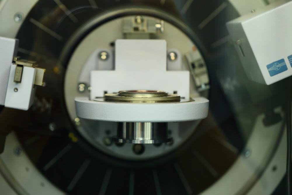
X-ray diffraction (XRD) is a non-invasive method for determining that can be used in phase analysis investigations of crystalline materials.

Image Credit: AgriTech/Shutterstock.com
The essential idea is that the constituent atoms in the crystal lattice cause an X-ray beam to scatter in a number of different directions—a change in the crystal size results in a variation of the width of the X-ray diffraction pattern.
Introduction to X-ray Diffraction
X-ray diffraction is a process in which the atoms of crystals generate a diffraction pattern of the waveforms existing in an incoming stream of X rays due to their equal intervals. The molecular dimensions of the crystal operate on the X rays in the same way as an evenly governed diffraction acts on a light beam.
On June 8, 1912, a thirty-three-year-old scientist named Max von Laue reported his findings of X-ray dispersion in the crystalline lattice as in a three-dimensional beam splitter at the German Physical Society conference at the University of Berlin.
The finding was utilized by two English researchers, W. H. Bragg and W. L. Bragg, father, and son, to validate Barlow's hypothesized conception of rock salt, launching the first X-ray diffraction examination of nanocrystals.
In recent times, in the field of nanotechnology, X-ray diffraction is regarded as a versatile method for characterizing the size of nanocrystals, which exhibits the capability of interacting with the electrons at an atomic scale.
Complimentary Techniques: XRD and XRF
The key difference between an X-ray fluorescence (XRF) analyzer and an X-ray diffraction (XRD) analyzer is that the former gives the nanocrystal sample’s elemental composition and the latter gives the sample’s chemical and structural information.
XRF is an analytical method in which the sample is excited by a primary x-ray source and the emitted fluorescent X-ray determines the elemental composition that quantifies the sample’s chemistry without deteriorating it. The sample’s elements emit a collection of fluorescent x-rays, which is specific to each unique element.
Hand-held models of XRF equipment give on-site, instant feedback regarding the elemental composition, which is useful for nanocrystal synthesis’ quality and process control. Since XRF analyzers fail to quantify the chemical and structural composition of nanocrystal samples, such composition can be quantified through XRD analyzers.
XRD analyzers scrutinize either the nanocrystal sample’s diffraction pattern or its characteristic X-ray scattering – both of which correlate to the atomic level information of the sample. XRD pattern comparison of an unidentified matter with that of a known one allows quantification of the unidentified matter’s properties.
XRD and XRF can be used as complementary analytical methods, even if both methods operate on different principles. For example, for a sample of CdS nanocrystals, XRF and XRD give the concentration and phase profile, respectively. A combined XRD-XRF analyzer comprises several benefits ranging from a diverse materials’ elemental analysis to phase determination of polyphase nanocrystalline materials.
Latest Research Findings
The field of nanotechnology and nanoscience extensively uses quantum dots, such as nanocrystals of Cadmium Sulfide (CdS) in various applications. For example, CdS nanocrystals have been shown to demonstrate two coexistent crystal structures: the polymorph of cubic zinc blend and the hexagonal structure of wurtzite. In this study, the team wrote that below the size of 10 nm, the peaks of the wurtzite overlap with that of the polymorph blend, and the resulting XRD patterns have intensities seemingly larger than the patterns of the wurtzite, making it too difficult to distinguish.
Many researchers have proved the method appropriate for analyzing the samples of nanocrystals containing mixtures of different particle sizes. According to Londoño-Restrepo, et al (2019), the XRD peaks of nanocrystals are found be broadened as their size decreases up to the nanoscale and further.
Urade, (2022) wrote that ideally, wurtzite cobalt sulfide (CoS) nanocrystals of average size 5 nm and 13 nm, respectively show the respective widths of the (002) peak and the (100) peak, which is mutually close but distinctive from one another in the XRD graph.
The latest research published in Nature scientific reports focuses on assessing the variation of the size of nanocrystals on the results of XRD patterns from humans and porcine bones.
TEM analysis revealed that polycrystalline nanocrystals with stretched plate shapes were essential and pristine constituents of raw bones. The findings validated the nano-dimensions of unprocessed bone hydroxyapatite, which are straight plate-like structures that might be associated with the bone's well-known transverse and selective growth.
The SEM analysis indicated that as with H-720, HAp crystals achieved diameters in the range of micrometer after the carbonization process at 720°C. Crystals with extended morphologies were found in the B-720 sample, and their borders were connected to those of other crystalline substances. It was proof that the coalescence-based growth mechanism had been disrupted.
A distinctive XRD pattern of a decorticated bovine bone revealed that a pattern is created by the following factors: specimen composition (polycrystalline and unstructured), disturbance (noise), and experimental functionality. However, it is crucial to demonstrate that the region utilized to define the crystallographic input of a nanocrystal specimen is created by elastic and plastic dispersion and the experimental component, implying that each of these signals cannot be determined individually. The XRD signals for raw HAps were characterized as polycrystalline specimens with "poor crystallographic integrity," yet the HRTEM pictures show that these samples belong to organized HAp nanocrystals.
Limitations and Challenges
The sensitivity of XRD results with the size of nanocrystals can be advantages as well as limitations. Researchers reported that even the decrease in size from 50 to 25 nm causes dramatic peak broadening, which further increases significantly for the crystal with a size less than 10 nm. Such nanocrystal sizes have low peak intensity, which makes it challenging to identify them as they are often found overlapped with one another.
Taking the example of HAp again, one of the issues with interpreting its XRD analysis spectra is the utilization of the FWHM as a metric to assess their crystalline nature. However, direct evaluation of this characteristic in the control sample reveals wide peaks.
BIO-HAp has been claimed to have inadequate crystalline quality based on the FWHM of a distinctive peak, and the crystallinity % is calculated using the whole XRD pattern. However, the large peaks shown in the diffraction patterns are caused by scattering from the BIO-HAp nanocrystals, widening caused by scattering, and experimental impact.
In short, the crystal size determines the form and amplitude of the X-ray diffraction peaks for structured crystalline material. The large peaks of the nano HAP patterns are not always associated with disorderly crystalline formations.
From the standpoint of the future of synthetic biology, these results demonstrated that the combustion procedure might be used to create biocompatible-HAp, though XRD studies must demonstrate that the temperatures did not cause alterations in its nano size.
Continue reading: Developing a Universal Route to Controlled Nanocrystal Synthesis
References
Nath, D., Singh, F., & Das, R. (2020). X-ray diffraction analysis by Williamson-Hall, Halder-Wagner and size-strain plot methods of CdSe nanoparticles- a comparative study. Materials Chemistry and Physics. 239. 122021. doi: 10.1016/j.matchemphys.2019.122021
Reischig, P., & Ludwig, W. (2020). Three-dimensional reconstruction of intragranular strain and orientation in polycrystals by near-field X-ray diffraction. Current Opinion in Solid State and Materials Science. 24(5). 100851. doi: 10.1016/j.cossms.2020.100851
Zhang, L., Gonçalves, A. A., & Jaroniec, M. (2020). Identification of preferentially exposed crystal facets by X-ray diffraction. RSC Advances, 5585-5589. 10(1). doi: 10.1039/d0ra00769b
Ooi, C.Y., Hamdi, M., and Ramesh, S. (2007) Properties of Hydroxyapatite produced by annealing of bovine bone. Ceramics International, 1171-1177. 33(7). doi10.1016/j.ceramint.2006.04.001
Holder, C., & Schaak, R. (2019). Tutorial on Powder X-ray Diffraction for Characterizing Nanoscale Materials. ACS Nano. doi:10.1021/acsnano.9b05157
Londoño-Restrepo, S., Jeronimo-Cruz, R., Millán-Malo, B., Rivera-Muñoz, E., & Rodriguez-García, M. (2019). Effect of the Nano Crystal Size on the X-ray Diffraction Patterns of Biogenic Hydroxyapatite from Human, Bovine, and Porcine Bones. scientific reports. doi:10.1038/s41598-019-42269-9
Mourdikoudis, S., Pallares, R., & Thanh, N. (2018). Characterization techniques for nanoparticles: comparison and complementarity upon studying nanoparticle properties. Nanoscale. doi:10.1039/C8NR02278J
Ravansari, R., Wilson, S., & Tighe, M. (2020). Portable X-ray fluorescence for environmental assessment of soils: Not just a point and shoot method. Environment International. doi:10.1016/j.envint.2019.105250
Urade, A. (2022). How is X-Ray Diffraction Used In Nanoanalysis? [Online] AZoNano. Available at: https://www.azonano.com/article.aspx?ArticleID=5955
Disclaimer: The views expressed here are those of the author expressed in their private capacity and do not necessarily represent the views of AZoM.com Limited T/A AZoNetwork the owner and operator of this website. This disclaimer forms part of the Terms and conditions of use of this website.