Mar 26 2009
In the last few years live cell imaging applications have increased dramatically, led by an array of new fluorochrome molecules with outstanding quantum efficiency and stability. However, capturing fast cellular processes without damaging the cells under observation and maintaining the optimum conditions for survival over a long period is still a challenge.

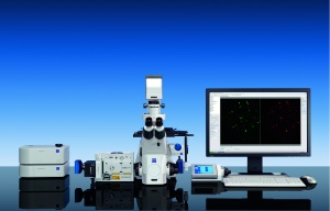
Carl Zeiss has responded with the launch of the Cell Observer SD, which fully integrates the CSU-X1 confocal scanning unit manufactured by Yokogawa Electric Corporation (Japan) and, for the first time, optimises the unit's features for the exacting requirements of live cell imaging. With Cell Observer SD, confocal observation and documentation of experiments on living cells over a long period of time and with high frame rates is possible.
"The combination of high resolution, sensitivity and speed is essential to track the communication and interaction of cells, organisms and structures," says Aubrey Lambert, Carl Zeiss UK. "Cell Observer SD makes that possible to deliver outstanding image quality and exceptional sensitivity and open up a new time window for confocal microscopy.”
Cell Observer SD is ideal for research in molecular cell biology, developmental biology, neurobiology and live cell imaging in general. Together with the entire line of incubation accessories from Carl Zeiss, Cell Observer SD enables users to observe living specimens for hours without damaging them. All major incubation parameters such as temperature and CO2 content are saved automatically together with the image data and all settings are also made via the AxioVision software.
The AxioVision software modules work solo or in combination, from simple camera control or multichannel imaging to a special high-speed mode for maximum frame rates or the simultaneous operation of two cameras. Other fluorescence imaging techniques, such as fluorescence recovery after photobleaching (FRAP), are also easily integrated.