May 4 2010
Electrical currents are invisible to the naked eye - at least they are when they flow through metal cables. In nerve cells, however, scientists are able to make electrical signals visible. Working with fellow experts from Switzerland and Japan, scientists from the Max Planck Institute for Medical Research in Heidelberg successfully used a specialized fluorescent protein to visualize electrical activity in neurons of living mice. In a milestone study, scientists are able to apply the method to watch activity in nerve cells during animal behaviour. (Frontiers in Neural Circuits, 29 April 2010)
Neurons communicate with one another via so-called action potentials. During an action potential, voltage-gated calcium channels are opened resulting in rapid calcium ion influx. Because of this tight coupling, fluorescent calcium indicator proteins can visualize action potentials. These proteins have two fluorescent subunits, one of which radiates yellow light and the other blue. When the proteins bind calcium, the proportion of yellow to blue light changes. Colour variation from blue light towards yellow thus reports different calcium levels - which is why the protein has been dubbed a "cameleon".
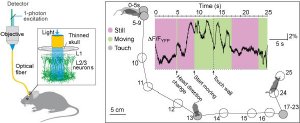
 Fibre-optic recording of YC3.60 signals in barrel cortex. Schematics of a setup (left) and functional signals in a freely-moving mouse (right). Image: Mazahir T. Hasan
Measuring action potentials optically
With the cameleon protein YC3.60, a fairly new variant, the scientists succeeded in recording the reaction of nerve cells to sensory stimuli in the intact brain of mice: every time the whiskers were deflected by a puff of air, there was a change of colour in the cameleon proteins in the nerve cells of the sensory areas of the cortex. It could therefore be deduced that the affected cells had reacted to the stimulus with action potentials. "The cameleon protein YC3.60 gives us the ability to measure action potentials not only in brain slices, but also in the intact brain. The molecule reacts quickly and sensitively and also captures changes in calcium concentrations occurring in rapid sequence," explains Mazahir Hasan from the Max Planck Institute for Medical Research.
The scientists were able to investigate activity in single cells as well as in whole groups of nerve cells. "YC3.60 has therefore proven to be a suitable tool for studying nerve tissue at different levels: on the one hand, we can monitor the fluctuation of calcium to infer action potentials within nerve cells. And what is even more advantageous, we can simultaneously measure the activity of neural networks or entire brain regions," says Mazahir Hasan. Consequently, the next step the scientists want to do is to selectively introduce cameleon proteins into a specific cortical layer or into different types of nerve cells. "Then we may be in a position to understand how different nerve cells in brain circuits generate complex behaviours," states Mazahir Hasan hopefully.
Measuring without electrodes
Cameleon proteins could therefore revolutionize the study of electrical activity in the brain. To date, the only way scientists could do this is by inserting electrodes into the nerve tissue or the cells. This electrode technique is blind to cell identity and it damages the tissue. By contrast, the cameleon protein’s colour changes can be observed in a much less invasive procedure using glass fibres as light conductors or with the help of modern fluorescence microscopes - known as two-photon laser-scanning microscopes. Moreover, cameleon proteins can be formed by cells themselves provided a corresponding section of DNA has been inserted into the genome in advance. In the experiments conducted by the scientists, viruses served as the vehicle for smuggling the genetic information for the cameleon proteins into the nerve cells.
In two earlier studies, an international team of scientists headed by Mazahir Hasan were the first to demonstrate that similar genetic probes can successfully detect natural sensation (such as smell and touch) in the mammalian brain in the form of unique activity patterns (Hasan et al., 2004) and, more importantly, with single-cell, single-action-potential resolution (Wallace et al., 2008). In the current study, they have reached yet another major milestone as they demonstrate that the cameleon YC3.60 can be used to record activity from a large number of nerve cells during behaviour in freely moving mice. Additionally, it is well suited for recording activity from the same nerve cells in the same animals over a long time period and should help scientists to understand how network activity patterns form to code for different experiences and animal behaviour.
These new advances, using light to study the brain, provide us with a unique opportunity to investigate how memories are formed and lost and, furthermore, when and where nerve cell activity patterns become altered as in the case of aging and also in neurological diseases such as Alzheimer’s disease, Parkinson’s disease and schizophrenia.