Jul 12 2010
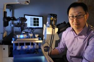
Chang Lu and his chemical engineering research group at Virginia Tech have discovered how to "greatly enhance" the delivery of DNA payloads into cells.
The description of their work will be featured on the cover of Lab on a Chip (issue 16), the premier journal for researchers in microfluidics.
 Chang Lu, an associate professor of chemical engineering at Virginia Tech, and his research group are featured in the July 8, 2010, issue of Nature and their work will also be in an upcoming issue of Lab on a Chip.
Lu's ultimate goal is to apply this technique to create genetically modified cells for cancer immunotherapy, stem cell therapy and tissue regeneration.
One of the most widely used physical methods to deliver genes into cells "is incredibly inefficient because only a small fraction of a cell's total membrane surface can be permeated," said Lu, an associate professor of chemical engineering at Virginia Tech.
The method Lu is referring to is called electroporation, a phenomenon known for decades that increases the permeability of a cell by applying an electric field to generate tiny pores in the membrane of cells.
Lu called the process "a new spin on DNA delivery." He explained the process saying, "Conventional electroporation methods deliver DNA only within a very small portion of the cell surface, determined by the physics governing the interaction between an electric field and a cell. Our method enables uniform DNA delivery over the entire cell surface, which is the first time we are aware that this has been demonstrated. The result is a greatly enhanced transfer of the genetic material."
Lu said his new approach harnesses "hydrodynamic effects that uniquely occur when fluids flow along curved paths. Flow under these conditions is known to generate vortices. Cells carried by such flow experience rotation and spinning that help expose the majority area of its surface to the electric field. " Having the gene delivery done by flows in curved paths is key in the gene delivery as opposed to the traditionally used, electroporation in static solution or in straight channels. "A spiral-shaped channel design yields a two-fold increase than a straight channel and an even larger factor compared to in static solution," he added.
By using fluorescence microscopy, they were able to "map" the area on the cell surface that was subjected to electroporation, and determine the extent of the DNA entry into the cell.
Lu explained the conventional delivery using a cuvette-type of device with static cell suspension produces DNA delivery confined to a narrow zone on the cell surface. However, when electroporation is applied to flowing cells in a spiral or curved channel , the images "appear dramatically different with the DNA delivery uniformly distributed over the entire cell surface."
Source: http://www.vtnews.vt.edu/