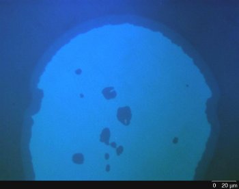
A research group from the Karlsruhe Institute of Technology (KIT) in Germany reports in Langmuir how SEEC microscopy can be used to provide a plausible scenario as to the behaviour of labelled lipids at a surface.
This new technique, based on the use of the new generation of microscope slides - referred to as Surfs - leads to the direct visualisation of nanometric samples through a conventional optical microscope. In addition, recent developments enable the complete topographic study of nanometric samples with a dedicated optical instrument.
 Lipids multilayer observed by SEEC microscopy.
Dip Pen Nanolithography (DPN) has been shown to be a powerful tool for the generation of biologically active surface patterns. Molecules are delivered from an AFM tip to a solid surface via capillary transport, making DPN a potentially useful tool for the elaboration of functionalised nanoscale devices. Due to the large covered area, systematic quality control of prepared patterns remains difficult. Atomic Force Microscopy (AFM) is not practical for extra large area screening while fluorescence microscopy (FM) can cause photo-chemical degradation and addition of fluorescent probes are needed that might have undesired side effects on the system.
Surface Enhanced Ellipsometric Contrast (SEEC) microscopy is carried out in combination with special microscope slides (Nanolane) that exhibit contrast-enhancing properties, which makes it possible to visualise nanometric structures (down to 0.3nm) with a 'standard' optical microscope. This technique requires absolutely no pre-treatment such as labelling. SEEC microscopy benefits from all optical microscopy features: real-time imaging; wide range of fields of view (from 70µm x 50µm to several mm²); the ability to perform studies in liquid to name but a few. In addition to that, some recent developments now allow for the conversion of a 2D SEEC image into a 3D map for complete topographic studies (profile extraction, step height measurements, roughness...). The accuracy of the measurements (0.3nm) is guaranteed thanks to certified calibration standards (from PTB Institute, Germany) and ISO 17025 procedures.
Recently, a Karlsruhe Institute of Technology (KIT) research group (Germany) led by Dr Hirtz has proposed a new structural model of lipid membrane stacking by combining information from 3 microscopic techniques including SEEC microscopy. Membrane stacks prepared with 20 mol % admixing of DNP Cap PE* to the DOPC** carrier are studied by AFM, FM and SEEC. Liss Rhod PE*** is used as a fluorophore molecule for FM analysis.
While AFM and SEEC topographic studies indicate three different levels of the membrane stack elaborated by DPN, thicknesses determined by AFM and SEEC conclude that the first layer is a monolayer whereas the second and third layers are bilayers. Surprisingly, FM does not display higher but lower fluorescence intensity for the third level. Fluorescence intensity drops almost to the level obtained for a single monolayer and is about 3.5 times lower than expected. Dr. Hirtz's group put forward two hypotheses to explain this phenomenon that occurs for a high-level of admixing of DNP Cap PE: (1) an enrichment of DNP CapPE in and under the third layer with decreased fluorescent intensity due to depleted Liss Rhod PE concentration or (2) an increase in Liss Rhod PE concentration in and under the third layer causing self-quenching of the fluorophore.
Scenario (1) is considered the most plausible. At room temperature, DNP Cap PE is in a gel state and DOPC and Liss Rhod are above their respective transition temperature, high amounts of admixing with DNP Cap PE should lead to phase separation of gel-state DNP Cap PE domains, while the fluid-state DOPC and Liss Rhod PE should be of better compatibility at room temperature.
Besides, comparing the optical thickness obtained by SEEC microscopy with the physical thickness from AFM measurements provides information about the refractive index of the different membrane stack layer. The refractive index of the first (mono)layer is determined to be higher than those of lipid bilayers (second and third layers). The rise of the layer compactness of a monolayer as compared to that of a bilayer appears to be a possible explanation for this result.
Although fluorescence microscopy can speed up the screening process after the lithographic process step, it may also lead to misinterpretations in the case of thin structures with high admixing of functional compounds as observed in this study. SEEC microscopy renders the use of fluorescent probe molecules pointless, which might come in handy for certain applications. Like AFM, SEEC microscopy is able to perform thickness measurements but without limitation for lateral screening. Combining both techniques is an elegant way to estimate the optical indices of unknown materials and gain information about some of their physical properties such as density.
The latest developments on SEEC microscopy are being conducted by Nanolane.
References:
*DNP Cap PE: 1,2-dipalmitoyl-sn-glycero-3-phos-phoethanolamine-N-[6-[(2,4-dinitrophenyl)amino]hexanoyl] )
**DOPC: 1,2-dioleoyl-sn-glycero-3-phosphocholine
***Liss Rhod PE: 1,2-dioleoyl-sn-glycero-3-phosphoethanolamine-N-(lissamine rhodamine B sulfonyl)