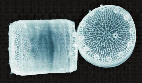
Scientists at the Pacific Northwest National Laboratory (PNNL) of the Department of Energy have developed a fluorescent biosensor using Thalassiosira pseudonana, a tiny marine diatom.
 A side and overhead view of the microscopic marine diatom Thalassiosira pseudonana. PNNL scientists used this species to develop a fluorescent biosensor that changes its glow in the presence of the sugar ribose. (Photo courtesy of Nils Kröger, Universität Regensburg.)
This biosensor paves the way to sense chemicals and other objects in water samples. It comprises fluorescent proteins embedded in the glassy shell of the diatom that glows while being exposed to a specific substance. This response will be helpful for scientists to synthesize a new class of diatom-inspired nanomaterials to solve issues in catalysis, sensing and environmental remediation.
Diatoms’ tiny shells, made of silica, are composed of complex, highly-ordered patterns. In the study, the PNNL researchers intended to utilize the fluorescent proteins to convert the diatoms into a reagent-free biosensor, which is capable of detecting a target object by itself and without the help of another substance or chemical. The research team introduced genes for its biosensor into the Thalassiosira pseudonana diatom, which has a hatbox-like shell. These new genes made the diatoms to generate a protein, which is the biosensor.
The biosensor’s core contains the ribose-binding protein, which can be attached to the sugar ribose. Every ribose-binding protein is then sandwiched by two other proteins, of which one glows in blue color and the second glows in yellow color. This three-protein complex is then bonded to the shell of the diatom as it grows.
When the ribose is not present, the two fluorescent proteins are placed close to each other and transfer energy through fluorescence resonance energy transfer, which makes the biosensor to emit both the colors. However, the ribose-binding protein alters the shape of the diatom when it is attached with the diatom, making the fluorescent proteins to be placed far apart from each other. This reduces the light energy emitted by the blue protein on the yellow protein, causing the biosensor to glow more in blue color.
The PNNL team measured the intensities of the specific light wavelengths emitted by the two fluorescent proteins using a fluorescence microscope that features a photon sensor. The team calculated the ratio of these two light wavelengths to identify the exposure of the diatom biosensor to ribose and the amount of ribose. The team was also able to run the biosensor with the shell alone, after its removal from the live diatom. This makes the silica biosensor more flexible and useful.