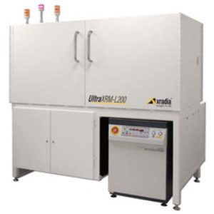
The Lawrence Livermore National Laboratory (LLNL) and Los Alamos National Laboratory (LANL) have each installed the UltraXRM-L200, Xradia’s lab-based microscope that delivers three-dimensional visualization at a resolution down to 50 nm.
 The UltraXRM-L200
With the installation of this 3D X-ray imaging solution, LLNL and LANL have further improved their capability for non-destructive material characterization. The advanced microscope delivers synchrotron-level resolution critical to 4D and in situ analyses of microstructural evolution, thus facilitating materials by design and other advanced research studies.
LLNL and LANL researchers conduct key material evolution and composition research for academia and government agencies. With the help of the UltraXRM-L200, they can now characterize dynamic microstructural properties under different conditions over time to understand biological advancements like joint replacements, sophisticated fusion methods utilized in advanced energy initiatives, design of materials utilized in global and national security.
The UltraXRM-L200 is the first-of-its-kind lab-based 3D X-ray microscope that provides ultra-high spatial resolution. With Zernike phase contrast imaging and Xradia's precision X-ray focusing optics, the microscope delivers consistent volumetric data down to 50 nm, which is otherwise possible only by cross-sectioning or by other destructive methods.
Scientists can take benefits from the in situ and 4D capabilities of the 3D X-ray microscope to explore microstructural changes with respect to time and subject specimens to different external conditions, including temperature, impact, and force, for evaluation of composition and deterioration. Design and evolution can be predicted and simulated accurately and efficiently by integrating physical imaging with computational assessments.