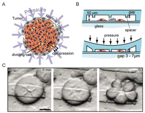
A group of bioengineers from the UCLA Henry Samueli School of Engineering and Applied Science has devised a microfluidic platform for the mechanical confinement of cancer cells to investigate the impact of three-dimensional (3-D) microenvironments on mammalian cell division or mitosis events.
 Cancer mitosis (credit: UCLA Engineering) Image Credit: (a) In vivo tumors are subjected to spatially and mechanically challenging conditions; (b) Profile view of microfluidic device; and (c) Cell division into five daughter cells
The microfluidic platform enabled for high-resolution, single-cell microscopic studies while the cells divided and grew. Unlike conventionally used culture flasks, this cutting-edge platform allowed the researchers to effectively simulate the in vivo environs of a cancer cell’s space-constrained development in 3-D environments.
What surprised the team during the study was such confinement made single cancer cells to divide abnormally into three or more daughter cells at a rate much higher than normal. Sometimes, a single cell divided into five daughter cells in a single division event, thus resulting in aneuploid daughter cells.
Principal investigator, Dino Di Carlo informed that albeit cancer can form from precise mutations, most malignant cancers have aneuploid cells and the cause for this is not yet clear. The novel microfluidic platform is a highly reliable tool for researchers to explore the role of distinctive tumor environment in aneuploidy.
According to the research team, the study of the contributing factors, which cause mismanagement during the chromosome segregation process, will help scientists better understand the development of cancer. The study results have been reported in the peer-reviewed PLoS ONE journal.
The UCLA Henry Samueli School of Engineering and Applied Science funded the study. At present, the research team is planning to team up with cancer researchers to conduct more research on the role of confined environments on the progression of cancer.