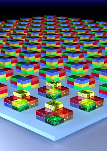
Borrowing from microfabrication techniques used in the semiconductor industry, MIT and Harvard Medical School (HMS) engineers have developed a simple and inexpensive way to create three-dimensional brain tissues in a lab dish.
 MIT engineers have devised a way to stack neurons to form three-dimensional brain tissue. (Image: Marcia Williams Medical Illustrations)
The new technique yields tissue constructs that closely mimic the cellular composition of those in the living brain, allowing scientists to study how neurons form connections and to predict how cells from individual patients might respond to different drugs. The work also paves the way for developing bioengineered implants to replace damaged tissue for organ systems, according to the researchers.
"We think that by bringing this kind of control and manipulation into neurobiology, we can investigate many different directions," says Utkan Demirci, an assistant professor in the Harvard-MIT Division of Health Sciences and Technology (HST).
Demirci and Ed Boyden, associate professor of biological engineering and brain and cognitive sciences at MIT's Media Lab and McGovern Institute, are senior authors of a paper describing the new technique, which appears in the Nov. 27 online edition of the journal Advanced Materials. The paper's lead author is Umut Gurkan, a postdoc at HST, Harvard Medical School and Brigham and Women's Hospital.
'Unique challenges'
Although researchers have had some success growing artificial tissues such as liver or kidney, "the brain presents some unique challenges," Boyden says. "One of the challenges is the incredible spatial heterogeneity. There are so many kinds of cells, and they have such intricate wiring."
Brain tissue includes many types of neurons, including inhibitory and excitatory neurons, as well as supportive cells such as glial cells. All of these cells occur at specific ratios and in specific locations.
To mimic this architectural complexity in their engineered tissues, the researchers embedded a mixture of brain cells taken from the primary cortex of rats into sheets of hydrogel. They also included components of the extracellular matrix, which provides structural support and helps regulate cell behavior.
Those sheets were then stacked in layers, which can be sealed together using light to crosslink hydrogels. By covering layers of gels with plastic photomasks of varying shapes, the researchers could control how much of the gel was exposed to light, thus controlling the 3-D shape of the multilayer tissue construct.
This type of photolithography is also used to build integrated circuits onto semiconductors — a process that requires a photomask aligner machine, which costs tens of thousands of dollars. However, the team developed a much less expensive way to assemble tissues using masks made from sheets of plastic, similar to overhead transparencies, held in place with alignment pins.
The tissue cubes can be made with a precision of 10 microns, comparable to the size of a single cell body. At the other end of the spectrum, the researchers are aiming to create a cubic millimeter of brain tissue with 100,000 cells and 900 million connections.
Answering fundamental questions
Because the tissues include a diverse repertoire of brain cells, occurring in the same ratios as they do in natural brain tissue, they could be used to study how neurons form the connections that allow them to communicate with each other.
"In the short term, there's a lot of fundamental questions you can answer about how cells interact with each other and respond to environmental cues," Boyden says.
As a first step, the researchers used these tissue constructs to study how a neuron's environment might constrain its growth. To do this, they placed single neurons in gel cubes of different sizes, then measured the cells' neurites, long extensions that neurons use to communicate with other cells. It turns out that under these conditions, neurons get "claustrophobic," Demirci says. "In small gels, they don't necessarily send out as long neurites as they would in a five-times-larger gel."
In the long term, the researchers hope to gain a better understanding of how to design tissue implants that could be used to replace damaged tissue in patients. Much research has been done in this area, but it has been difficult to figure out whether the new tissues are correctly wiring up with existing tissue and exchanging the right kinds of information.
Another long-term goal is using the tissues for personalized medicine. One day, doctors may be able to take cells from a patient with a neurological disorder and transform them into induced pluripotent stem cells, then induce these constructs to grow into neurons in a lab dish. By exposing these tissues to many possible drugs, "you might be able to figure out if a drug would benefit that person without having to spend years giving them lots of different drugs," Boyden says.