Mar 30 2013
David DeRosier is championing a new scientific method. Call it the retired-biology-professor-goes-back-to-the-lab method.
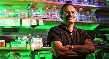
 Emeritus Professor of Biology David DeRosier
An emeritus professor of biology, DeRosier has been working as a postdoctoral fellow in neuroscientist Gina Turrigiano’s lab for several years. This job bookends DeRosier’s first postdoc in the famous Cambridge, England, lab where Watson and Crick discovered the structure of DNA, and numerous other Nobelists, including DeRosier’s mentor, Aaron Klug, also made fundamental biological discoveries.
With characteristic understatement, DeRosier says, “It was an exciting time.”
DeRosier might be the most decorated postdoc ever, considering his long-standing membership in the U.S. National Academy of Sciences and several other prestigious scientific societies. Now the Microscopy Society of America has honored him with its Distinguished Scientist Award in Biological Sciences.
The award recognizes “pre-eminent senior scientists from both the biological and physical disciplines who have a long-standing record of achievement during their career in the field of microscopy or microanalysis.”
In Turrigiano’s lab, DeRosier is drawing on this track record to build a super-resolution cryogenic light microscope. Earlier in his career, the biologist pioneered the use of electron microscopy to make fundamental discoveries about cellular structures called molecular machines.
Being a postdoc, says DeRosier, “is a way to give back to Brandeis and a different way to think about retirement. I am glad I picked Gina’s lab. She is incredibly smart; it makes her lab a great place to work.”
He and Turrigiano are collaborating to create a new tool for understanding a major focus of Turrigiano’s research: how synapses (structures that permit neurons to pass an electrical or chemical signal to another cell) undergo constant change in response to learning and development while maintaining stability.
“There are more than 1,000 different proteins in the synapse,” says DeRosier. “We can’t tag all of them for electron microscopy, so we are trying a different approach, using a super-resolution light microscope to examine the structures in a frozen hydrated state using a cold stage.”
“This is a project that we just could not have tried without someone like David to spearhead it,” says Turrigiano. “There was fierce competition among Brandeis neuroscience labs to recruit David for his second postdoc. It’s been great collaborating with him these past several years, and I feel incredibly lucky to have such a distinguished and brilliant postdoc in my lab.”
For his part, DeRosier says he’s enjoying his unconventional retirement: “I get to do science, and it’s what I enjoy. It’s humbling to walk into a new field and to be in the position of learning something new.”