Jul 25 2016
A team of international researchers working at Penn State have developed a highly sensitive chemical sensor based on Raman spectroscopy. This was developed using nitrogen-doped graphene as a substrate. In this scenario, doping introduces nitrogen atoms into the carbon structure of graphene.
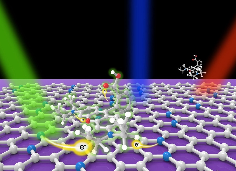
 A model showing the charge transfer (e-) mechanism of Rhodamine B molecules (top) interacting with N-doped graphene (bottom sheet) when excited with different laser lines, which leads to ultrasensitive molecular sensor with N doped graphene. The white, blue and red balls represent carbon, nitrogen and oxygen atom respectively. CREDIT: Terrones Lab / Penn State
This method helps to identify trace amounts of molecules present in a solution at extremely low concentrations, some 10,000 times more diluted than can be seen by the naked eye.
Raman spectroscopy is an identification technique that has been extensively used in materials science, chemistry, and the pharmaceutical industry to detect the unique internal vibrations of a wide variety of molecules. When molecules or crystals are irradiated by a laser light, colors are scattered and shifted. It is possible to detect the scattered light in the form of a Raman spectrum, which acts as a fingerprint for all Raman-active irradiated systems.
Basically, different colors in the visible spectrum will be associated to different energies. Imagine each molecule has a particular light color emission, sometimes yellow, sometimes green. That color is associated with a discrete energy.
Mauricio Terrones, Professor, Penn State
Three varieties of fluorescent dye molecules were chosen by the team for their experiments. Fluorescent dyes, which are often used as markers in biological experiments, are very difficult to detect in Raman spectroscopy because the fluorescence can wash out the signal. The photoluminescence - fluorescence - is quenched when the dye is added to the N-doped graphene substrate or the graphene.
The Raman signal is extremely weak on its own, so a number of techniques have been used to improve the signal. An enhancement technique that has been developed recently uses pristine graphene as a substrate, which has the potential to improve the Raman signal by several orders of magnitude.
The journal Science Advances published a paper on July 22, 2016 that presents a repot by Terrones and colleagues on the addition of nitrogen atoms to the pristine graphene to further enhance sensitivity. The researchers presented a theoretical explanation of how N-doped graphene and graphene cause the enhancement.
"By controlling nitrogen doping we can shift the energy gap of the graphene, and the shift creates a resonance effect that significantly enhances the molecule's vibrational Raman modes," said lead author Simin Feng, a graduate student in Terrones' group.
This is foundational research. It is hard to quantify the enhancement because it will be different for every material and color of light. But in some cases, we are going from zero to something we can detect for the first time. You can see a lot of features and study a lot of physics then. To me the most important aspect of this work is our understanding of the phenomenon. That will lead to improvements in the technique.
Ana Laura Elias, Research Associate, Penn State
Terrones added, "We carried out extensive theoretical and experimental work. We came up with an explanation of why nitrogen-doped graphene works much better than regular graphene. I think it's a breakthrough, because in our paper we explain the mechanism of detecting certain molecules."
The researchers hope that the newly developed method will effectively detect trace amounts of organic molecules because of graphene's chemical inertness and biocompatibility. Elias expressed her excitement at integrating the new method with the available portable Raman spectrometers that can be used in remote places to identify unsafe viruses, for example.
The researchers analyzed that the fluorescent dyes will allow fast and effortless detecting of compounds present inside biological cells. This simple method only requires the graphene substrate to be dipped into a solution for a short time period. Terrones stated that the method should be feasible to develop an entire library of the Raman spectrum of specific molecules.
Researchers from Japan, China and Brazil contributed to this work while visiting the Terrones lab at Penn State. The paper is titled "Ultrasensitive Molecular Sensor Using N-doped Graphene through Enhanced Raman Scattering."
Other coauthors include Maria Cristina dos Santos, Brazil; Bruno R. Carvalho, Brazil; Ruitao Lv, China; Qing Li, China; Kazunori Fujisawa, Yu Lei,Nestor Perea-López, all from Penn State; Morinobu Endo, Japan; Minghu Pan, China; and Marcos A. Pimenta, Brazil.
The research received MURI grants from the U.S. Army Research Office and the U.S. Air Force Office of Scientific Research. Agencies within their home countries extended support for the visiting faculty and students.