A team of researchers recently published a paper in the journal Nano Letters that demonstrated the effectiveness of fluorescent nanodiamond (FND)-based biocompatible nanoprobe to map the subwavelength features of tightly focused light fields under living conditions.

Study: Biocompatible Nanotomography of Tightly Focused Light. Image Credit: Heiti Paves/Shutterstock.com
Bioimaging and Optical Techniques
Tightly focused lights are one of the crucial tools to investigate the molecular and subcellular structures. However, the refractive index in biological tissues displays spatial heterogeneity, which often scatters the light and severely affects the actual focal field distribution.
Thorough knowledge of the focal field is necessary to ensure optimal performance in optical manipulation and imaging. Although different experimental techniques were previously developed to characterize the focused light fields, their applications in living systems faced several challenges such as photothermal effects and cytotoxicity.
FNDs possess ultralow cytotoxicity, improved cell absorption capacity, and a suitable functional surface for biomedical applications, and thus can be potentially used as an optical probe to characterize focal fields in living systems.
In this study, researchers selected the nitrogen-vacancy (NV) center in diamond to map the focused near-infrared (NIR) light field in biotissues. The working principle of the study was based on the sensitivity of NV center fluorescence to the NIR light intensity.
The Study
A 1064 nm NIR laser and 532 nm green laser were merged and tightly focused on both bulk diamond and FNDs placed on a three-dimensional (3D) piezo nano scanner. The NV center was excited by a green laser and the fluorescence (FL) was collected between 593 and 775 nm.
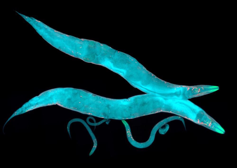
The FNDs were introduced in the digestive tracts of the Caenorhabditis elegans (C. elegans) by direct feeding to map the NIR focal field in the nematode. C. elegans typically display a robust autofluorescence background.
The charge modulation method was used to selectively extract the FND signal from the autofluorescence background as the method provides 80% absolute fluorescence contrast and works for both NV- and NV0 charge states. Additionally, the method can be effective for FNDs with sizes below 40 nm and can work at low NIR intensity.
Through this approach, a NIR light with a power density of 10 kW/cm2 can induce 6−22% fluorescence enhancement on FNDs.
A single FND was used on a coverslip as an optical probe to characterize the tightly focused optical field. The focal intensity landscapes obtained from FNDs in the C. elegans and on the coverslip were compared by using a tightly focused Gaussian beam.
Observations
Two stable charge states that include a negative NV- and a neutral NV0 were observed in the NV center in bulk diamond. The NV- charge state emitted a major part of the fluorescence upon excitation by NIR light.
The fluorescence emission was decreased when the NIR laser accelerated the ionization between NV0 and NV-. However, the fluorescence responded negatively to the NIR laser intensity when the high-intensity green laser was focused on the NV center.
No response was observed on the fluorescence emission by the NV center due to the NIR polarization rotation, indicating that the fluorescence contrast was determined by the local NIR intensity and the ionization process was not dependent on the NIR polarization. This polarization independence is specifically beneficial for biological applications where the nanoprobes randomly rotate in biotissues.
The NIR intensity profile suitably matched to a Gaussian distribution, where two significantly lower peaks were observed on both sides of the central Gaussian peak. Two-dimensional (2D) scanning demonstrated that these lower side peaks corresponded to the Airy disk.
FNDs with a group of NVs displayed a higher fluorescence compared to the bulk diamond with single NV centers. FNDs were also absorbed by cells without displaying any noticeable toxicity, indicating that functionalized FNDs can be effectively delivered to specific organelle and cells.
The FNDs submerged in the high autofluorescence background of C. elegans when they were excited with a green laser, even after the use of confocal imaging to suppress the fluorescence from the out-of-focus region. However, the background was separated and an enhanced signal-to-background performance was obtained when the NIR pulse sequence was applied to specifically modulate the FND fluorescence.
The FNDs were trapped and moved several hundred nanometers in the digestive tract of the nematode using the NIR beam as an optical tweezer. However, the FNDs jumped back to their initial positions when the moving distance became much larger, indicating their adherence to the surface cells.
The focus diameter in the C. elegans was larger compared to the coverslip due to the spherical aberration incorporated by the difference in the refractive index of C. elegans and coverslip. The biotissue scattering introduced certain random disturbances to the light phase when the Bessel beam was tightly focused in the C. elegans.
The photon shot-noise was the primary error source in the intensity measurement. The contrast measurement error was 0.3% with a green laser power of 1.3 μW and a sampling time of 0.2 s.
To summarize, the findings of this study demonstrated that FNDs can effectively provide light field characterization for optical tweezers and super-resolution imaging as a biocompatible nanoprobe under living conditions.
Reference
Zhang, Q., Yin, J., Yan, Y. et al. (2022) Biocompatible Nanotomography of Tightly Focused Light. Nano Letters https://pubs.acs.org/doi/10.1021/acs.nanolett.1c03905
Disclaimer: The views expressed here are those of the author expressed in their private capacity and do not necessarily represent the views of AZoM.com Limited T/A AZoNetwork the owner and operator of this website. This disclaimer forms part of the Terms and conditions of use of this website.