Color-enzyme-based lateral flow immunoassay (LFIA) can amplify the color signal after catalyzing the substrate. In an article published in the journal Analytical Chemistry, researchers applied vanadium disulfide nanosheet (VS2NS) as LFIA enzyme label to detect 17β-estradiol (E2) and enhance LFIA tests.

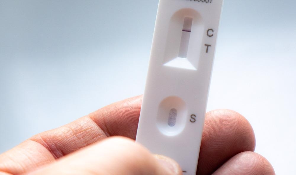
Study: Vanadium Disulfide Nanosheet Boosts Optical Signal Brightness as a Superior Enzyme Label to Improve the Sensitivity of Lateral Flow Immunoassay. Image Credit: Ian Gessey/Shutterstock.com
They observed a more prominent enzyme catalytic performance in VS2NS than natural horseradish peroxidase (HRP).
Nanomaterials in LFIA
LFIA is widely used in the food sector, medical diagnostics, genotype analysis, and environmental monitoring. Although colloidal nanoparticles (NPs) are widely used in LFIAs, more exploration is required to cope with the need for rapid detection and signal amplification. To this end, enzyme-catalyzed LFIA provides great potential in expanding the detection range and amplifying the color signal. However, natural enzymes do not have a color, consequently, they require a nanocarrier to serve as a signal label.
Recently layered transition-metal dichalcogenide (TMD) materials have gained considerable interest in the nonlinear optical field due to their broadband gap, ultrafast carrier dynamics, and third-order optical nonlinear polarization. TMD can also serve as two-dimensional (2D) nanozymes due to their unique surface area and exposed active sites, which improves their catalytic activity. VS2NS is a common TMD material with peroxidase-like activity and higher adsorbing ability than HRP.
Dual Signal LFIA for E2 Detection
In the present study, the researchers addressed bottleneck issues in natural enzyme-based LFIAs. They introduced VS2NS as an enzyme label to obtain a dual signal LFIA. E2 is extensively used in poultry breeding and livestock and its residual amounts in the environment enter the food chain, ultimately affecting the male reproductive system.
In this work, the researchers chose E2 as a model analyte to realize an accurate detection. Moreover, VS2NS with all favorable properties was anticipated to bring about high performance as LFIA enzyme label. The aggregation of VS2NS-labeled probes induced a distinct optical signal due to their movement towards immobilized antigen on the test (T) line. Furthermore, adding 3,3’,5,5’-tetramethylbenzidine (TMB)−hydrogen peroxide (H2O2) solution induced the peroxidase-like activity in VS2NS, which led to the oxidation of substrate TMB and production of blue enzyme catalysis signals.
Research Findings
The VS2NS was prepared via a modified solvothermal method. The team used a green liquid exfoliation method to fabricate an ultra-thin VS2NS. The scanning electron microscopy (SEM) images revealed that the precursor, VS2·NH3 had a laminar morphology, and was exfoliated into VS2NS of size less than 1 micrometer after subjecting to ultrasound for half an hour and a size of fewer than 200 nanometers after subjecting to ultrasound for 2 hours, suggesting the suitability of VS2NS for the capillary force. Tapping mode atomic force microscopy (AFM) revealed the thickness of VS2NS, which was less than 5 nanometers.
The X-ray diffraction (XRD) spectrum obtained for VS2NS showed characteristic peaks that coincided with standard VS2NS. The ultraviolet-visible (UV-vis) absorption showed that VS2NS oxidized TMB into a blue oxidation product with an absorption peak at 650 nanometers in the presence of hydrogen peroxide (H2O2). Contrastingly, TMB was unoxidized in the absence of VS2NS, as inferred from the solution that remained colorless, suggesting the peroxidase-like activity of VS2NS. The VS2NS-catalyzed decomposition of H2O2 results in the generation of hydroxyl free radical (•OH) that oxidizes the TMB substrate to a blue-colored product. The researchers proved that VS2NS with low dimensions exhibited superior catalytic activity over bulk VS2. The enhanced catalytic activity of VS2NS is attributed to reduced screening effects in monolayered to few-layered structures that lead to enhanced Coulomb interactions.
E2-BSA captured the VS2-monoclonal antibody (mAb) probes, after adding them to the solution, resulting in the formation of black color on T line, which was derived from VS2-mAb probes captured by the T-line due to specific interactions between E2-BSA and antibodies. However, the addition of VS2NS did not show the black color in the LFIA. VS2NS-mAb probes were formed by modifying the purified anti-E2 mAb onto the surface of VS2NS via electrostatic interactions.
The Fourier-transform infrared (FTIR) spectra of the VS2NS showed characteristic peaks between 400-1000-centimeter inverse and the peaks between 500−690 and at 978- centimeter inverse corroborates V-S-V and V-S stretching vibrations. Moreover, the peaks corresponding to amide I and amide II of the antibody polypeptide chain appeared at 1500 and 1700-centimeter inverse, respectively.
In summary, the researchers exploited a few-layered VS2NS-based LFIA with strong adsorbing capacity and a large surface area to volume ratio. These properties of VS2NS brightened the signal and enhanced the detection sensitivity. Thus, the superior peroxidase-like activity of VS2NS helps amplify the label signal.
Reference
Chen, Y., Ren, J., Yin, X., Li, Y., Shu, R., Wang, J. and Zhang, D., (2022) Vanadium Disulfide Nanosheet Boosts Optical Signal Brightness as a Superior Enzyme Label to Improve the Sensitivity of Lateral Flow Immunoassay. Analytical Chemistry,. https://pubs.acs.org/doi/10.1021/acs.analchem.2c01008
Disclaimer: The views expressed here are those of the author expressed in their private capacity and do not necessarily represent the views of AZoM.com Limited T/A AZoNetwork the owner and operator of this website. This disclaimer forms part of the Terms and conditions of use of this website.