Chitin is a renewable biopolymer that is abundantly available in nature. Despite its importance as a basic building block in various biological materials, its surface crystalline structure remains unexplored.

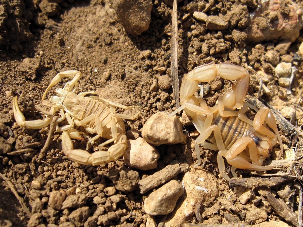
Study: Probing the Structural Details of Chitin Nanocrystal–Water Interfaces by Three-Dimensional Atomic Force Microscopy. Image Credit: Malpolon/Shutterstock.com
In an article recently published in the journal Small Methods, the researchers employed atomic force microscopy (AFM) and molecular dynamics (MD) simulations to reveal the structural details of the chitin nanocrystal (chitin NC)-water interface at the molecular level.
Composition of Chitin
β (1-4) linked N-acetyl-d-glucosamine residues are the building blocks in the chitin chain that are commonly found on crustaceans’ shells. Chitin has many structural and functional similarities with cellulose. Chitins like cellulose are found in nature in the form of ordered crystalline microfibrils along with a few proteins and minerals. Pure chitin NCs are obtained from crab and shrimp shells via mechanical and chemical treatments. Depending on the source, chitin exists in three types of polymorphs, α, β, and γ chitins. However, the most stable form is α-chitin.
The morphological and structural details and chemistry of chitin determine its properties. However, the molecular-scale details on chitin NC’s surface structure at its interface in an aqueous medium are limited, and it is imperative to understand the three-dimensional (3D) structural organization of water molecules at its interface on chitin’s surface to determine the kinetics of enzymatic hydrolysis.
Structural Details of Chitin NC–Water Interface
In the present work, the chitin NCs were isolated from shells of shrimp in the water, and their structural details at the molecular level were realized by combining MD simulations with frequency modulation 3D atomic force microscopy (FM-3D-AFM). The structural details of individual NC’s chitin chain arrangements were realized from a highly resolved AFM image, which showed a well-ordered surface without many structural defects. The 3D-interfacial hydration layers of structured water molecules on the chitin NC surface were visualized with subnanometer resolution at the single-chain level.
On the chitin NC surface, the water molecules are stable molecularly ordered hydration-layered structures, which encapsulate the chitin NC surface inhomogeneously. This suggests that the chitin NC’s amphiphilic surface character consists of multiple crystalline planes, different chain arrangements, and the ability to form hydrogen bonds.
MD simulations aided in corroborating these findings and the results aligned with those obtained from AFM. The obtained results can serve a crucial purpose in exploring the enzymatic and chemical degradation at the aqueous chitin interface and understanding the structure-property relationships at chitin NC surfaces.
Research Findings
The chitin NCs were prepared from chitin powder via hydrochloric acid hydrolysis, and their morphology and size distribution were assessed by transmission electron microscope (TEM) and FM-AFM measurements.
The TEM images of chitin NCs showed needle-shaped nanostructures of 10-20 nanometer diameter and 100-600 nanometer length on chitin fragments. Further, the images obtained from AFM confirmed the homogenous morphologies with laterally aggregated and isolated NCs. The less-charged surface or incomplete fibrillation of NCs resulted in the formation of chitin NC aggregates.
The diameter of individual NC was observed to be 10 to 40 nanometers and 100-300 nanometers in length. The observed diameter of chitin NCs was larger than their actual size due to tip-convolution effects. Moreover, the histogram analysis performed on different samples confirmed that the height distribution was 4-15 nanometers which is consistent with the chitin NC’s average size, derived from shrimps.
The FM-AFM images revealed that the individual chitin NC’s surface had an extreme chain ordering that covered the entire crystal surface. The observed high degree of crystallinity was free of any structural defects, indicating that the amorphous domains are mainly composed of proteins.
The minerals were removed through acid treatment while retaining the crystalline structures. Structural characterization of different individual chitin NC surfaces at the molecular level confirmed that the structural features at the interface are best accessed when the axis of chitin NC is parallel to the probe or at an angle of 45 degrees.
At the solid-liquid interface, the AFM tip interacts with surface features and an ordered layer of solvent molecules on the surface. The MD simulations and AFM measurements confirmed that the hydration structures observed are equivalent to the underlying structures of chitin NCs at the molecular level.
The upper half of the AFM image revealed a zigzag arrangement of chitin chains, and the image’s lower half displayed a row-like pattern. The surface functional groups with different chain orientations resulted in two types of surface arrangement in chitin NCs. Additionally, the researcher hypothesized that water molecules might rearrange the interface to maximize the interaction with the surface of chitin NC. High-resolution AFM showed different contrast patterns due to the hydrogen bonds formed by the exposed amide and hydroxyl groups with the neighboring water molecules.
In conclusion, the researchers characterized the chitin NC surface at the atomic and molecular levels. They anticipate that the research outcomes can help gain a deeper understanding of chitin NC’s surface chemistry and structure-activity relationships. The present study could pave a path to assess the enzymatic and chemical activities on the surface of chitin NCs.
Reference
Yurtsever, A., Wang, P.-X., Priante, F., Morais, Y., Miyata, K., MacLachlan, M. J., Foster, A. S., Fukuma, T., (2022) Probing the Structural Details of Chitin Nanocrystal–Water Interfaces by Three-Dimensional Atomic Force Microscopy. Small Methods, 2200320.https://onlinelibrary.wiley.com/doi/full/10.1002/smtd.202200320
Disclaimer: The views expressed here are those of the author expressed in their private capacity and do not necessarily represent the views of AZoM.com Limited T/A AZoNetwork the owner and operator of this website. This disclaimer forms part of the Terms and conditions of use of this website.